Neural tube defects: Nursing
Introduction0:00–0:19
Neural tube defects or NTD for short, refer to developmental defects of the central nervous system secondary to failed closure of the neural tube during the fetus's embryonic development.
Now let's quickly look at the physiology of neural tube development. Let's zoom in on a cross section of the early embryo.
Physiology0:19–2:52
The first one located internally gives rise to the neural tube. The second one located externally gives rise to skin cells and the epidermis.
And the third population called the neural crest cells develop in between the neural tube and epidermis and give rise to various structures throughout the body.
The mesoderm on the other hand, develops into a transient midline structure called the notochord, which produces various signals that guide the development of other embryonic structures with respect to the midline.
During the 3rd and 4th weeks of development, the notochord signals for the formation of the neural tube through a process called neurulation which is highly dependent on adequate folic acid levels.
Neurulation progresses in two stages. Primary neurulation begins when the ectoderm right above the notochord thickens and gives rise to the neural plate.
At both ends of the neural plate, there are neural crest cells and beyond them, the ectoderm that gives rise to the epidermis.
Soon after the edges of the neural plate thicken and tilt upward forming the so called neural folds. This allows for au shaped neural groove to form which sets the limit between the right and left sides of the embryo.
During secondary neurulation, the neural folds eventually fuse at the midline forming the hollow neural tube separate from the epidermis above it.
Now, let's look at a different view of the developing embryo this time from above at this point like any hollow tube. The neural tube still has openings at both ends, a large opening at the top and called the cranial neuropore and a smaller opening at the bottom end called the caudal neuropore.
The cranial neuropore seals up around day 25 while the caudal neuropore seals up a few days later, around day 28 the neural tube eventually develops into the brain and spinal cord while the notochord gives rise to a part of the intervertebral disc called the nucleus pulposus.
The cause of neural tube defects is not known. However, they are associated with any factor that can interfere with the development of the central nervous system during embryonic development.
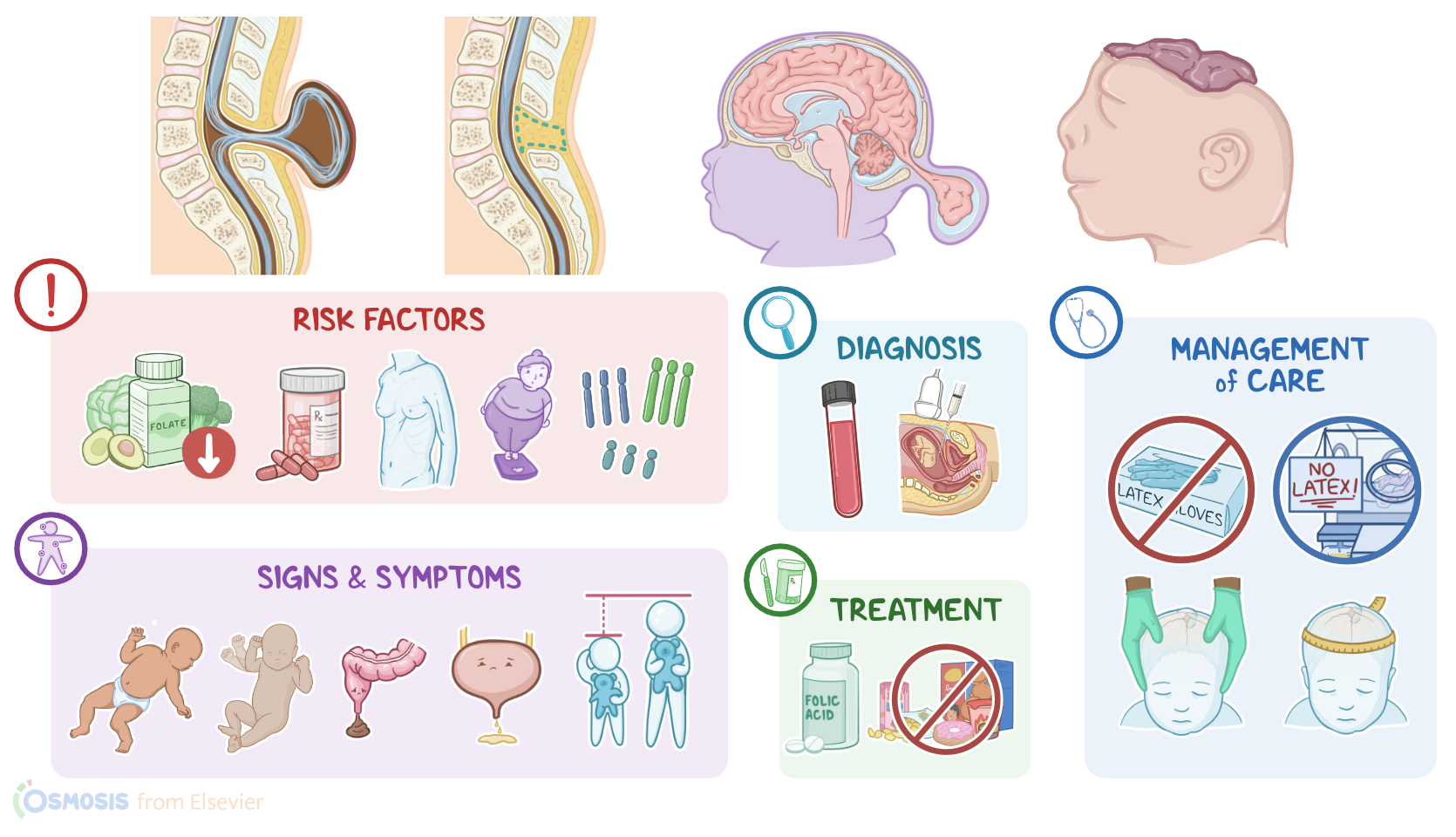
These include modifiable risk factors such as folate deficiency malnutrition, maternal obesity infections, medications such as sodium valproate and methotrexate and certain chemicals.
Causes & risk factors2:52–3:35
There are also non modifiable risk factors such as advanced maternal age, maternal diabetes and fetal genetic abnormalities including trisomy, 1318 and 21 pathology wise.
One or more of these risk factors increase the chance of failed neural plate folding or closure during neuralation. More specifically, this can occur either at the cranial neuropore involving the brain and skull or at the caudal neuropore involving the spinal cord.
Now, given the two main locations of neural tube defects, there are two types of defects that can occur. First, spinal defects are more common and can be further divided into two subtypes, spina bifida, occulta is where the posterior vertebral arches fail to close in the lumbosacral area.
Pathology3:35–6:11
However, in this case, the spinal cord and the meninges remain in their place and the defect is not usually visible from the outside.
Hence the name occulta, then spina bifida cystica is when parts of the spinal cord herniate through the abnormal gap in the vertebrae forming a cyst.
A meningocele is when only the meninges herniate into the cyst. While a myelomeningocele contains the meninges and spinal cord.
Second, cranial defects are mostly represented by encephalocele and it is when the brain and the meninges protrude through a defect of the skull.
Another cranial defect is anencephaly which is where the infant has small or missing brain hemispheres, upper skull and scalp secondary to failed closure of the cranial neuropore.
These infants are typically born without both a forebrain and a cerebrum and the remaining brain tissue may be exposed. Now, large defects come with a series of complications of their own.
For example, a large encephalocele allows the structures in the region to herniate. And if the defect is not corrected and blood flow compromised, it can cause significant damage to the nervous system.
Subsequently, central nervous injury early in the development of the embryo can result in cognitive delay, poor motor skills or cerebral palsy.
After birth. In some cases, severe neural tube defects can also cause spontaneous abortion or death in utero hydrocephalus or build up of fluid in the brain ventricles, polyhydramnios and the chiari malformation.
A chiari malformation is where the lower part of the brain herniates down into the spinal canal. Sometimes a spinal NTD can fistulate causing cerebrospinal fluid to leak out through the skin, increasing the likelihood of a central nervous system infection.
Clinical manifestations of neural tube defects vary depending on the type of defect. Now, spina bifida occulta is usually invisible and asymptomatic.
If the defect is visible, it is usually seen only as a dimple on the back which may be covered in hair, spina bifida, cystica is when the same defect is visible as a protrusion or cyst of the meninges through the vertebrae with meningocele, the spinal cord remains in the correct position and these infants often present with minor or no neurologic deficits.
Clinical manifestations6:11–8:22
However, a myelomeningocele has a high risk of neurological deficits because the spinal cord herniates out of the vertebral defect, making it easier to damage.
Thus, increasing the risk of developing motor and sensory dysfunction below the defect. Additionally, the infant with such a defect has an increased risk of meningitis, hemorrhage and hypoxia.
Hence, the infant might present with symptoms, suggestive of these complications with a myelomeningocele, bladder and bowel incontinence can also be present.
Next. An encephalocele is seen as a round protrusion in the occipital area.
Generally, the protrusion is covered by skin, but it can also be open depending on its location and size. An encephalocele can cause cognitive deficits as well as neurological issues like spastic quadriplegia ataxia or a of muscle control or coordination of voluntary movements and visual problems.
Encephaloceles are also accompanied by craniofacial abnormalities like microcephaly and by severe cognitive deficits such as developmental delay and intellectual disability.
But some Children can achieve standard I QS. Finally, with anencephaly, infants are typically born without both a forebrain and a cerebrum and the remaining brain tissue might be exposed and it's incompatible with life.
Prenatal diagnosis of neural tube defects is usually done by testing the amniotic fluid via amniocentesis and maternal blood for increased levels of alpha fetoprotein or AFP and acetylcholinesterase at week 16 and 18 AFP is a fetal protein produced by the liver and leaks from the fetus into the amniotic fluid through exposed capillaries of the NTD acetylcholinesterase.
Diagnosis8:22–9:31
On the other hand, leaks directly from exposed neural tissue into the amniotic fluid. In cases where AFP levels are increased, high resolution fetal ultrasonography or fetal MRI can be used to diagnose neural tube defects prenatally after birth diagnosis can be established by physical examination and imaging such as CT and MRI.
Additionally, a cranial ultrasound can help diagnose some neural tube defects and assess for associated complications such as hydrocephalus.
Let's first look at the prevention of neural tube defects. One of the most efficient ways to prevent a neural tube defect is by ensuring folic acid supplementation.
Individuals who plan to give birth should consume 400 mcg of folic acid daily for around six months prior to conception, increasing the dose to 600 mcg daily during pregnancy.
Additionally, addressing the risk factors predisposing to neural tube defects such as obesity through weight loss, diabetes through strict glycemic control and discontinuation of drug use will also reduce the risk of a defect occurring.
Treatment9:31–11:32
Now, if a neural tube defect is detected during prenatal screening, parental counseling regarding the prognosis should be offered and there should be a conversation around the decision to continue the pregnancy.
If pregnancy is continued, delivery should be planned and the optimal delivery mode is usually by Cesarean section at a facility where intensive neonatal care can be provided.
In addition, in cases with the myelomeningocele, fetal surgery could be attempted to stop the leakage of cerebrospinal fluid which can reduce the risk of chiari malformation and hydrocephalus.
After birth management depends on the type and severity of the defect. For example, those with spina bifida, occulta do not require immediate medical intervention.
More severe defects such as encephalocele and myelomeningocele usually require surgical repair in order to place the displaced tissues back into the skull or spinal canal and to remove the meningeal sac where possible if hydrocephalus is present, a ventriculo peritoneal shunt placement might be needed for drainage of the cerebrospinal fluid.
A shunt is a thin tube that's surgically implanted in the brain and drains away the excess fluid surgery to correct the defect is recommended 72 hours after delivery.
Ok. Let's look at the care you'll be providing for a newborn with myelomeningocele.
Your priority goals are to minimize heat and fluid loss, prevent complications and provide psychosocial support immediately after birth.
Minimize heat and fluid loss from the spinal defect by covering it with a warm sterile saline soaked dressing and by placing the newborn feed first into a sterile bowel bag, then place the infant in a radiant warmer or Isolette with temperature control and monitor their temperature closely.
Remember to position them prone or on their side in order to avoid putting pressure on the defect and to prevent tearing the membrane that's covering the defect.
Management and care11:32–14:28
Then infuse the prescribed IV fluids to replace insensible fluid losses and place an indwelling urinary catheter to monitor fluid output.
Also be sure to take steps to prevent complications because infants with spinal defects are at increased risk of developing a latex allergy.
Be sure to use latex free gloves and other supplies during care and post assign by the infants bedside alerting all caregivers to only use latex free supplies.
Since the infant is at an increased risk for infection, administer the prescribed IV antibiotics. Also remember that infants with myelomeningocele often have fecal incontinence.
So be sure to keep stool off of the defect and prevent skin breakdown by providing scrupulous skin care to the perineal area.
Because infants with a myelomeningocele often develop hydrocephalus, palpate the cranial sutures to look for separation and fontanelles to check for pressure.
Also measure their occipital frontal head circumference to determine if it's within normal limits during care. You'll also want to decrease environmental stimuli by reducing light and noise and by clustering activities as much as possible to allow periods of rest.
Assess the infant frequently for signs of pain using an appropriate pain scale like the neonatal pain, agitation and sedation scale or NPAS and administer the prescribed analgesics.
Once the infant is stable, assist with preparing for surgical closure of the defect. Now keep in mind that this is a time of high stress and anxiety for the infant's family.
Be sure to encourage them to voice their fears and concerns and involve them in the care of their baby whenever possible.
Let them know you understand how overwhelming it can be to have a child with a serious medical condition and continue to provide emotional support as well as counseling services as needed.
Ok. Moving on to client and family teaching begin by explaining that myelomeningocele is a defect where the neural tube fails to close during development.
So part of their baby's spinal cord protrudes out of the back forming a sac. Be sure to provide clear explanations about the care their baby is receiving.
Answer any questions they have and reassure them that a team of specialists will be part of their baby's treatment when their baby is ready to go home.
Talk to them about how their baby will be supported by home health and an early intervention program to enhance their baby's physical cognitive language and social development.
Stress the importance of keeping all scheduled appointments to ensure the best outcome for their baby. Be sure to instruct them to seek medical attention right away if their baby develops new symptoms such as lethargy, choking or trouble swallowing, squeaky, or labored breathing changes in their voice, worsening, bowel and bladder problems or signs of a seizure, including jerking or twitching movements, excessive blinking or prolonged staring.
General client and family teaching14:28–16:08
Lastly emphasize the importance of consulting with their health care provider if they plan on additional pregnancies. So they can be supported with nutrition, folic acid supplements, optimal weight gain and blood glucose control as needed as well as prenatal care and monitoring.
All right, as a quick recap. Neural tube defects are central nervous system defects that occur when the neural tube fails to close during embryonic development.
The exact cause isn't known but theyre associated with risk factors including folate deficiency malnutrition, infections, medications like sodium valproate and methotrexate.
Advanced maternal age diabetes and obesity. N TDS are also associated with fetal genetic abnormalities including trisomy.
1318 and 21 types of N TDS include spina bifida, occulta spina bifida, cystica, myelomeningocele encephalocele and anencephaly.
Clinical manifestations of NTD S can include absent motor or sensory function below the defect, orthopedic deformities, bladder and bowel incontinence and developmental delay diagnosis can be done prenatally by testing the amniotic fluid and maternal blood for a FP and acetylcholinesterase.
Review16:08–17:03
After birth diagnosis is done by physical examination and imaging as well as cranial ultrasound that can assess for complications like hydrocephalus.
Importantly, prevention of NTD is done with folic acid supplementation and addressing modifiable risk factors. Optimal delivery is often through cesarean section and treatment involves surgical repair of the defect.
Priority goals of nursing care include minimizing heat and fluid loss, preventing complications and providing psychosocial support.
Client and family. Teaching focuses on care of the baby at home when to seek medical attention and prevention of NTD S in future pregnancies.
| NEURAL TUBE DEFECTS (NTDs) | ||
| KEY POINTS | NOTES | |
| DEFINITION |
| |
| PHYSIOLOGY |
| |
| CAUSES AND RISK FACTORS |
| |
| PATHOPHYSIOLOGY |
| |
| SIGNS AND SYMPTOMS |
| |
| DIAGNOSIS |
| |
| TREATMENT |
| |
| MANAGEMENT OF CARE |
| |
| PATIENT AND FAMILY TEACHING |
| |
No notes for this video yet
Try adding a note below