Physiologic changes - Postpartum: Nursing
Introduction0:00–0:22
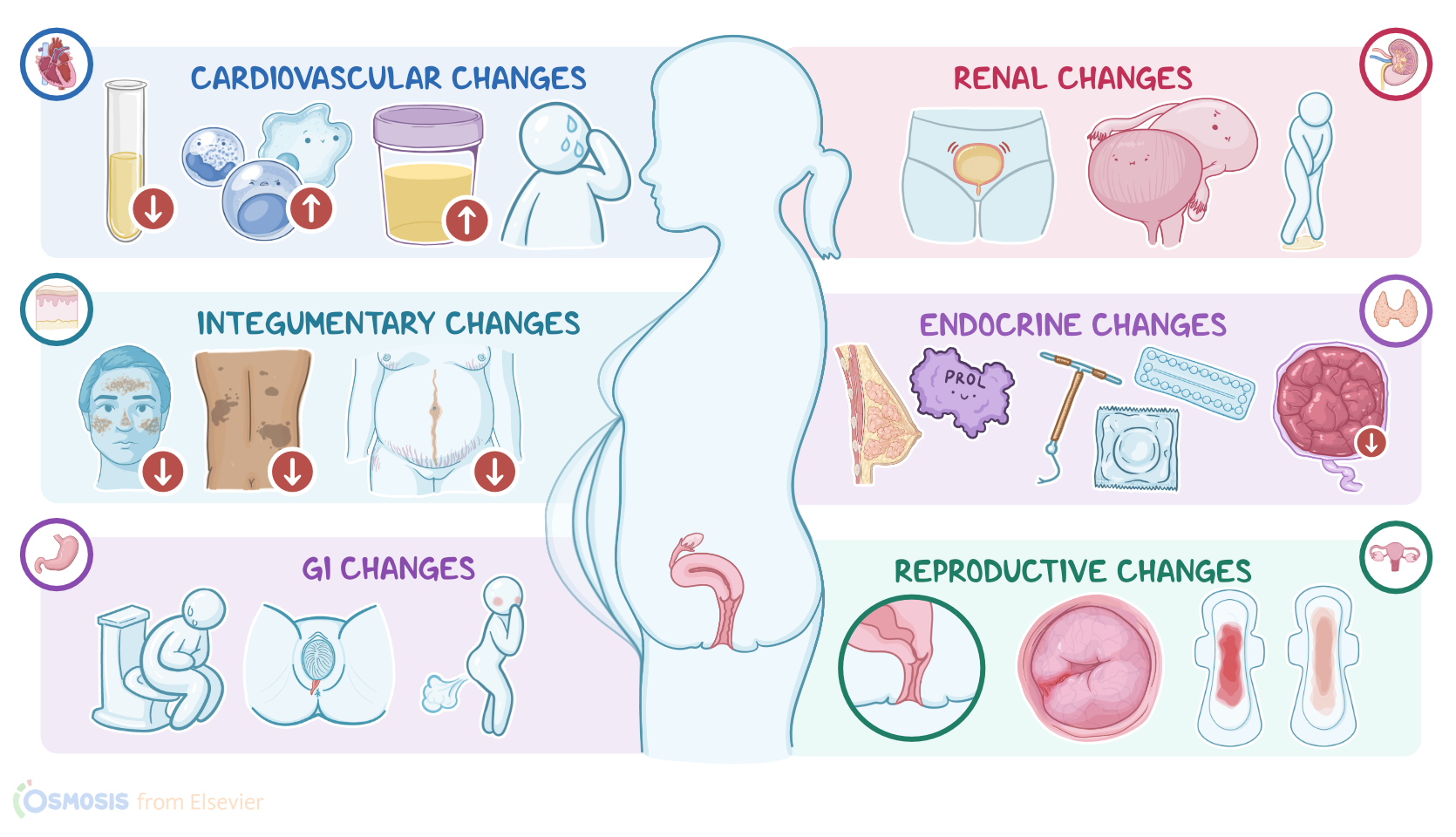
The postpartum period, also known as puerperium, is defined as the first six weeks after delivery. Physiologic changes during the postpartum period include the reversal of changes that occurred during pregnancy.
Moreover, these changes are primarily caused by a rapid drop in estrogen and progesterone. Now, after delivery, a client usually loses 4500 to 5800 grams or 10 to 13 lb, which covers the weight of the fetus, amniotic fluid, and the placenta.
Weight loss0:22–0:48
The weight loss starts immediately after delivery and continues over the next several months due to normalization of blood volume and increased caloric expenditure from milk production.
Okay, let’s focus on reproductive changes, starting with the uterus. After delivery, the uterus begins to return to its nonpregnant state of size and position, a process called uterine involution.
Reproductive changes0:48–4:23
As soon as the placenta is delivered, uterine muscle fibers constrict uterine blood vessels, preventing a life-threatening condition called postpartum hemorrhage.
Uterine contractions, often referred to as afterpains because they cause sharp pain in the lower abdomen, continue during the postpartum period to further aid uterine involution.
After that, it normally descends by about 1 centimeter, or 1 fingerbreadth, per day, until it reaches the pelvic cavity by the 14th day.
Now, let’s take a look at vaginal discharge after birth, called lochia. There are three types; lochia rubra, lochia serosa, and lochia alba.
Lochia rubra refers to the dark red vaginal discharge that is present for the first 3 days. It consists of blood, small blood clots, decidua, and mucus.
As the bleeding reduces, the volume of vaginal discharge reduces, and lochia rubra transforms into lochia serosa. Lochia serosa refers to the thin, red to brown vaginal discharge that lasts until the 10th day after delivery.
It consists of white blood cells, serous exudate, and cervical mucus. As time passes, lochia serosa transforms into lochia alba.Lochia alba refers to the yellowish-white vaginal discharge that typically lasts until the 14th day after delivery, but can persist up to 6 weeks.
Lochia alba is rich in epithelial cells, white blood cells, fat, and bacteria. Additionally, it’s important to assess the amount of lochia on the perineal pad every 60 minutes.
After delivery, the internal os of the cervix fully closes, while the external os remains slightly open at about 2-3 centimeters at 2 to 3 days post-delivery.
By the end of the first week, it narrows to less than 1 centimeter, and appears as a transverse slit. The baby also causes trauma to the vaginal walls, which results in edema and lacerations.
After delivery, the vagina loses folds and ridges of the vaginal walls called vaginal rugae, but they start to reappear 3 to 4 weeks after delivery.
Cardiovascular & hematological changes4:23–5:54
At 6 weeks after delivery, the vagina reaches a near pre-pregnant size.Now, moving on to cardiovascular changes. After delivery, the blood that used to supply the uterus returns to systemic circulation, causing a transient increase in cardiac output that eventually returns to the prelabor values one hour after delivery.
Plasma volume also decreases after delivery. First, there’s a normal delivery related blood loss, which is typically 200 to 500 mL for a vaginal delivery ,and 600 to 800 mL for a cesarean delivery.
Also, aldosterone and oxytocin production decreases, thereby increasing diuresis and fluid loss through urine; and there’s increased diaphoresis or sweating.
Together, these processes decrease the plasma volume and return it to the prepregnancy levels by 6 weeks postpartum. Moreover, this also normalizes the hematocrit values and coagulation factor levels.
This usually occurs over the next 4 to 6 weeks. However, during this period, clients remain in a hypercoagulable state and are at increased risk for thromboembolic events Finally, during the first 24 hours, the white blood cell count increases up to 30,000 cells per mm3.
Gastrointestinal changes5:54–6:31
This increase in white blood cell count is a physiologic response and is not associated with an active infection. Usually, it takes around one week for the body to normalize the white blood cell count.
Next up are gastrointestinal changes. After delivery, the most common problem is constipation often accompanied by flatulence and abdominal fullness.
The first reason this can occur is because progesterone levels from pregnancy remain elevated right for several days after delivery and this hormone reduces gastrointestinal tone and motility.
Renal changes6:31–7:55
Also, if a client had an episiotomy or perineal laceration during delivery, they might avoid defecation because of pain and discomfort.
Normal gastrointestinal motility usually returns at 2 to 3 days post-delivery. Let’s switch gears and look at renal changes during the postpartum period.
The kidneys typically return to their normal position over the next 4 weeks, while diuresis fully restores within 12 hours after delivery.
But, on some occasions, a client can develop urinary retention, which is an inability to empty all the urine from the bladder.
This can be caused by loss of bladder tone and elasticity, which can result in overdistension and subsequent retention of urine; alternatively, it could be a result of the pressure exerted by the fetus during delivery which decreases sensation in the urinary tract; or from trauma to the bladder, urethra, or urinary meatus.
Other causes of urinary retention include some medications, anesthesia, and lack of privacy. Urinary retention can interfere with uterine involution, and can often be recognized by an elevated or laterally displaced uterus.
It also provides an environment for bacterial growth and the development of urinary tract infections. Finally, some clients might experience stress urinary incontinence, which refers to involuntary urine leakage during exertion, such as laughing, coughing, and sneezing.
Endocrine changes7:55–8:48
This usually occurs due to trauma to the pelvic floor muscles and the bladder sphincter. Clients usually resolve stress urinary incontinence with exercises that strengthen the pelvic floor muscles.Next up are endocrine changes.
After delivery of the placenta, placental hormones, such as estrogen, progesterone, human chorionic gonadotropin, and human placental lactogen begin to decrease.
On the other hand, the anterior pituitary gland continues prolactin secretion, which triggers milk production and prevents ovulation.
Now, since prolactin inhibits ovulation, it causes lactational amenorrhea, meaning that it delays ovulation and menstruation while the mother is still breastfeeding.
Integumentary changes8:48–9:14
Breastfeeding clients can take 10 weeks to 6 months to start ovulating and menstruating again, but this only takes 6 to 10 weeks for non-breastfeeding clients.
Musculoskeletal changes9:14–9:55
However, both breastfeeding and non-breastfeeding clients should consider the use of contraceptive methods to prevent closely spaced pregnancies.
Next up are integumentary changes during the postpartum period. Hyperpigmentations and cutaneous vascular changes, such as spider angioma, telangiectasia, and palmar erythema fade away after delivery.
On the other hand, purple to red stretch marks called striae gravidarum fade away to a white-ish shade, but never fully disappear.
Neurological changes9:55–10:11
Now, switching gears and moving on to the musculoskeletal system. During labor, muscles are under increased stress.
This can result in muscle fatigue that primarily affects the neck, shoulders, and arms. Additionally, the hormone relaxin, which loosens pelvic ligaments and joints in preparation for labor, disappears.
Review10:11–11:25
Finally, after delivery, the uterus relieves pressure on the abdominal wall and resolves the previous separation of muscles called diastasis recti.
But, still, some clients might require additional abdominal exercises to fully restore the muscle tone of the abdominal wall and return it to the prelabor state.
Finally, let’s cover the neurological changes during the postpartum period. These primarily include headaches, which usually occur due to changes in fluid and electrolyte balance.
Also, regional anesthesia and dural punctures from spinal anesthesia can cause severe headaches. Alright, as a quick recap… Physiologic changes during the postpartum period include the reversal of changes that occurred during pregnancy.
After delivery, the uterus returns to its nonpregnant state in
| PHYSIOLOGIC CHANGES - POSTPARTUM | ||
| KEY POINTS | NOTES | |
| DEFINITION |
| |
| WEIGHT LOSS |
| |
| REPRODUCTIVE CHANGES |
| |
| CARDIOVASCULAR & HEMOTOLOGICAL CHANGES |
| |
| GASTROINTESTINAL CHANGES |
| |
| RENAL CHANGES |
| |
| ENDROCRINE CHANGES |
| |
| INTEGUMENTARY CHANGES |
| |
| MUSCULOSKELETAL CHANGES |
| |
| NEUROLOGICAL CHANGES |
| |
No notes for this video yet
Try adding a note below