Definitions & Key takeaways

Acute kidney injury (AKI) is a sudden, potentially reversible decline in renal function. AKI results in the accumulation of water, nitrogenous wastes, sodium, and other metabolic wastes in the body, and can also lead to electrolyte imbalances. Common causes include dehydration, sepsis, and some drug therapy.

AKI often presents with reduced urine output, but there can be other signs such as edema, and those associated with the accumulation of metabolic wastes such as anorexia, seizures, and altered mental status. The nursing process for AKI begins with the assessment and identification of patients at risk for developing the condition. Once AKI is diagnosed, nurses work with other health care providers to implement therapies aimed at preventing progression and maximizing patient outcomes. If AKI is left untreated, it can lead to chronic kidney disease (CKD).

Chapters:

Client Report0:00–1:36

Maurice Jones, a 58-year-old black man, sustained a back injury several weeks ago while cleaning out his garage, and since then he's been taking ibuprofen 3 to 4 times each day for pain.
Mr. Jones has a history of hypertension for which he takes the angiotensin 2 receptor blocker or ARB losartan, though he says sometimes he forgets to take it.
He also takes the proton pump inhibitor or PPI omeprazole for gastrointestinal reflux disease or GERD. He presented to his primary care provider or PCP with fatigue, headache, lower extremity edema, and decreased urine output.
His vital signs were oral temperature 99.0 °F or 37 °C, heart rate 98 BPM, respiratory rate 20 breaths per minute, and BP 148/96 millimeters of mercury.
Labs were ordered which showed BUN 28 mg per deciliter, creatinine 2.4 mg per deciliter, sodium 124 milliequivalents per liter, and potassium 5.3 milliequivalents per liter.
Arterial blood gas analysis revealed pH 7.25, partial pressure of carbon dioxide 42, partial pressure of oxygen 85, and bicarbonate 20.
Urine specific gravity is 1.010. Mr.
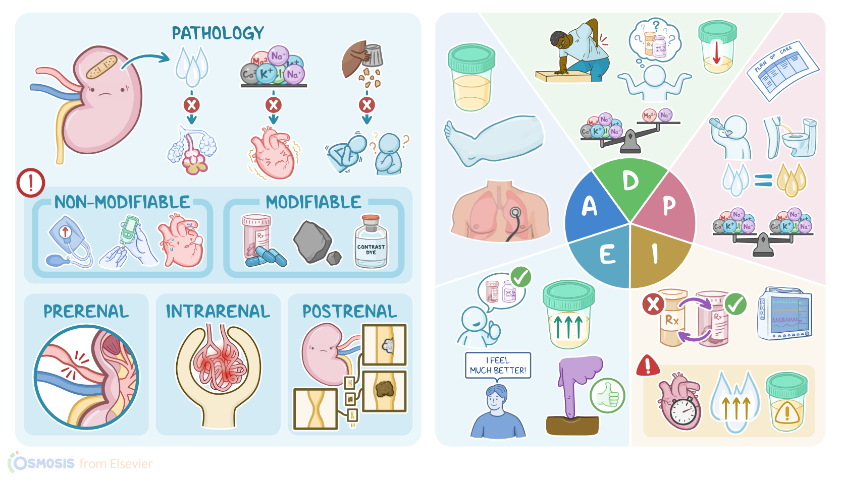
Jones was admitted to the medical unit with a diagnosis of acute kidney injury or AKI. Acute kidney injury, or AKI for short, refers to a sudden decrease in kidney function over hours or days.

Pathophysiology1:36–3:30

As a result, the kidney's ability to manage fluid, electrolyte, and acid-base balance is impaired, and there's decreased excretion of waste products such as urea and creatinine, which build up in the blood.
There are some factors that can put an individual at risk for AKI. Unfortunately, many of them are non-modifiable factors that decrease kidney function over time, such as advanced age, diabetes mellitus, autoimmune diseases, certain cancers, uncontrolled hypertension, and heart, liver, or kidney disease.
Some modifiable risk factors include exposure to nephrotoxic agents such as certain antibiotics, heavy metals, or chemotherapy, or being exposed to iodinated IV contrast dye during radiology procedures.
Now, the causes of AKI can be classified into pre-renal, intrarenal, and post-renal. In pre-renal AKI there is no physical problem with the kidneys themselves, but the problem is caused by decreased blood flow to the kidneys.
This can happen when there's a severe loss of body fluid due to problems like hemorrhage, burns, or ascites. Other common causes involve decreased cardiac output like with heart failure, myocardial infarction, or shock.
Intrarenal AKI occurs due to damage to the kidney itself. Some common causes include inflammatory problems like glomerulonephritis and damage done by toxins or prolonged ischemic injury.
Finally, postrenal AKI occurs due to decreased outflow of urine from the kidneys. This can be a result of something compressing the ureter like tumors, strictures, or an enlarged prostate, or from an obstruction caused by kidney stones lodged in the ureters.

Clinical Features and Complications3:30–4:46

AKI generally progresses through three phases oliguric, diuretic, and recovery. Often the first sign of AKI is decreased urine output, which can lead to fluid overload, peripheral edema, and weight gain, as well as hypertension.
A bounding pulse and distended neck veins could also be present. Over time, this can lead to complications like pulmonary edema, which causes dyspnea and tissue hypoxia.
Next, electrolytes can also become imbalanced since the damaged kidneys aren't able to excrete them effectively from the body.
Those who develop hyperkalemia may experience muscle weakness and dangerous cardiac arrhythmias. In addition, phosphate retention leads to hyperphosphatemia, which can ultimately lead to hypocalcemia resulting in tetany, which are involuntary muscle cramps.
As uremic waste products build up in blood, the client can experience symptoms like anorexia or appetite loss, nausea and vomiting, as well as uremic encephalopathy, which is initially characterized by confusion and lethargy and may progress to seizures and even coma.
Finally, if AKI is not treated, it may progress to permanent kidney damage or end-stage renal disease and death. The main diagnostic studies used for AKI include standard laboratory tests that can show abnormal electrolyte levels like hyperkalemia, hyperphosphatemia, and hypocalcemia.

Diagnosis and treatment4:46–6:01

Blood urea nitrogen or BUN and creatinine will also be elevated. A blood gas analysis can show metabolic acidosis.
In addition, urinalysis can reveal proteinuria, hematuria, and casts. Finally, imaging techniques like an ultrasound or CT scan can assess renal blood flow and allow identification of an underlying cause.
The good news is that AKI can be reversible when promptly treated, so the goal is to prevent further injury, manage the signs and symptoms, prevent complications, and promote recovery.
The underlying cause should be addressed while carefully managing the body's fluids, electrolytes, and waste products of metabolism.
This is achieved via a combination of IV fluids, diuretics, and nutritional support. Those who don't respond to treatment may require renal replacement therapy such as dialysis to control their volume overload, electrolyte imbalances, metabolic acidosis, and uremic symptoms.

Assessment6:01–7:25

As you enter the room to meet Mr. Jones, you perform hand hygiene, introduce yourself as his nurse, confirm his identity, and begin his assessment.
On his physical exam, Mr. Jones's lungs are clear, and he has no jugular venous distention or JVD.
You note 2 + pedal and ankle edema bilaterally. There is 250 mL of clear yellow urine in his urinal, and a bedside bladder scan measures 45 mL of retained urine.
Vital signs are oral temperature 99.6 °F or 37.5 °C. Heart rate 94 BPM and regular, respiratory rate 16 breaths per minute.
BP 144/90 millimeters of mercury, oxygen saturation 94% on room air, and pain 5 out of 10. He weighs 195 lbs or 88.5 kg and is 76 inches tall.
His morning labs include BUN 30 mg per deciliter, creatinine 2.6 mg per deciliter, sodium 128 milliequivalents per liter, potassium 5.5 millequivalents per liter, urinalysis shows the presence of casts, increased sodium, and a specific gravity of 1.010.

Nursing diagnoses7:25–7:43

EKG shows normal sinus rhythm. Based on the assessment data you collected, your nursing diagnoses include impaired fluid balance related to renal insufficiency and decreased urine output, electrolyte imbalance related to renal insufficiency, pain related to back injury, and deficient knowledge related to medication self-care.

Planning7:43–8:03

Next, you collaborate with Mr. Jones and the interdisciplinary team to plan goals to promote his optimal health.
Goals for Mr. Jones before discharge include balanced intake and output, normal electrolyte levels, and that he will verbalize understanding of medication self-care.

Implementation8:03–9:31

OK, now you are ready to implement your plan. The physician has replaced Mr.
Jones's losartan with the calcium channel blocker amlodipine and the loop diuretic furosemide temporarily to help mobilize fluid.
Acetaminophen is prescribed for pain, and for GERD, the H2 receptor antagonist cymetidine is prescribed. You administer these medications as ordered.
Next, you assign the certified nursing assistant or CNA the tasks of keeping strict INO's and daily weights. You continue to monitor Mister Jones's heart rhythm until his electrolyte levels are normalized.
As you interact with Mister Jones throughout the shift, you monitor him closely and will alert the attending physician if there is an abnormal change in his heart rhythm, if there is a significant change in his fluid balance, or if his laboratory values indicate his condition is worsening.
Next, you teach Mr. Jones about the potential effect of hypertension on his kidneys and the importance of taking his BP medication as directed to keep his BP within a normal range.
You explain how certain medications like NSAIDs and PPIs can impair kidney function, especially when taken together for an extended period of time.
You let him know he has a referral for physical therapy to help address his back pain. Then you document your ongoing care and teaching as you prepare to give report to the oncoming shift.

Evaluation9:31–10:09

The next morning, you are happy your evaluation shows Mr. Jones's urine output has increased.
Edema in his lower extremities is +1. His morning lab values include BUN 25 mg per deciliter, creatinine is 1.8 mg per deciliter, sodium 130 milliequivalents per liter, and potassium 5.1 millequivalents per liter.
Mr. Jones says he is feeling much better and he's looking forward to starting physical therapy after discharge.
He tells you he understands that he needs to adhere to his medication regimen to avoid injury to his kidneys. All right.

Summary10:09–11:10

As a quick recap, you have been caring for Mr. Jones, who was admitted to the hospital with AKI, which is a sudden decrease in kidney function that impairs the body's ability to excrete waste products, such as urea and creatinine, and can result in altered fluid, electrolyte, and acid-base balance.
Your nursing assessment reveals Mr. Jones has fatigue, oliguria, and lower extremity edema.
The nursing diagnoses include impaired fluid imbalance, electrolyte imbalance, and deficient knowledge. The plan of care for Mr.
Jones is to balance intake, output, and electrolytes, and to increase his understanding of how certain medications can affect his kidney function.
Together with the interdisciplinary team, you implement interventions to help him achieve these goals, and you will continue to evaluate his response and revise the care plan as needed to achieve optimal outcomes.