Shock - Septic: Nursing
Introduction0:00–0:31
Shock is a life-threatening condition that occurs when body organs don’t receive enough oxygen and nutrients for them to function properly.
Shock can be grouped into four types based on the cause: hypovolemic, cardiogenic, obstructive, and distributive shock. And septic shock is a type of distributive shock that occurs secondary to sepsis, which is when the body mounts an excessive inflammatory response because of an infection.
Now, to understand distributive shock, let’s quickly review the physiology of blood vessels, which contain smooth muscle in their walls.
Normal physiology0:31–1:50
When the smooth muscle relaxes, it increases the diameter of blood vessels, called vasodilation. On the other hand, when smooth muscle contracts, that decreases the diameter of blood vessels, called vasoconstriction.
The contraction and relaxation of smooth muscle is primarily controlled by the sympathetic nervous system, which normally maintains a partial constriction, generating enough force to keep blood moving through the circulatory system.
The sympathetic system stimulation can increase or decrease to keep up with the body’s needs. Increased sympathetic stimulation of the blood vessels causes vasoconstriction, which leads to increased mean arterial pressure, or MAP for short.
Vasoconstriction, when the total blood volume is constant, raises blood pressure and allows blood to flow faster through the blood vessels.
In contrast, vasodilation is typically caused by decreased sympathetic stimulation, in addition to inflammatory cytokines and histamine, which decrease mean arterial pressure.
So, vasodilatation, when blood volume is constant, lowers the blood pressure and slows down the blood flow through the blood vessels.Alright, now the main cause of septic shock is infections, particularly those caused by gram positive bacteria, like Staphylococcus aureus, Streptococcus pneumoniae or Enterococcus species, and gram-negative bacteria, like Escherichia coli.
Causes & risk factors1:50–2:56
In addition to bacteria, infections caused by viruses, fungi, or parasites can also cause septic shock. Typically, risk factors for developing septic shock include clients who are immunocompromised.
This includes clients who are in the extremes of age, such as children and the elderly, as well as in clients with diabetes, chronic kidney or liver disease, as well as clients with an underlying HIV infection, those with chronic glucocorticoid treatment, and those with cancer who are being treated with chemotherapy.
Other risk factors include prolonged hospitalization, indwelling catheters, as well as major surgery, and large open wounds.Alright, now the pathological process of septic shock starts when a pathogen, say a gram-negative bacteria, enters the body and finds its way into the bloodstream.
Pathology2:56–6:51
Embedded within the outer wall of this bacteria, there are types of lipopolysaccharides called endotoxins. These endotoxins are released when the Gram negative bacteria are destroyed by the immune system, which activates immune cells.
In turn, immune cells release proinflammatory cytokines, like interleukins and tumor necrosis factor, or TNF for short. These proinflammatory cytokines trigger excessive and widespread inflammation, called systemic inflammatory response syndrome, or SIRS.
This response causes increased production and release of stress hormones, like catecholamines and glucocorticoids, as well as a response in the hypothalamus, blood vessel walls, as well as the capillary endothelial cells.
On the other hand, some of these proinflammatory cytokines act as pyrogens. This means that they act on the hypothalamus to raise core body temperature above the normal set-point, which causes a fever.
Next, the vessel walls start producing nitric oxide, or NO for short, which is a vasodilator. Finally, the capillary endothelial cells refer to a decreased response to vasoconstricting stimulation, as well as a breakdown of the endothelial barrier, which increases permeability of blood vessels.
This causes fluid to move from the vessels into the interstitial space, resulting in systemic hypotension. In addition, the endothelial cells attract more platelets, which stick into the endothelial cells, forming tiny clots called microvascular thrombi.
All of these changes in SIRS decrease tissue perfusion, which causes tissue hypoxia. Now, the next events in septic shock are grouped into four stages: initial, compensatory, progressive, and refractory stages.
In the initial stage, systemic vasodilation causes less blood to return to the heart through the venous system; this, in turn, decreases cardiac output.
As a consequence, body cells are not well-perfused, so they switch to anaerobic metabolism to produce energy, causing lactic acid to build up in the blood.
When lactic acid builds up, that lowers blood pH, causing metabolic acidosis. This activates the compensatory stage.
During the compensatory stage, the sympathetic nervous system increases its activity to compensate for the decreased tissue perfusion in order to maintain homeostasis, and acid-base compensatory mechanisms activate to correct the acidosis.
In the progressive stage, these compensatory mechanisms start failing , so the body’s metabolic needs can’t be met. This results in lactic acid buildup in the blood, more than in the initial stage, and gradually worsening metabolic acidosis.
Finally, the refractory stage is when shock progresses to cellular death and multiple organ damage, which can be fatal. Complications of septic shock include acute respiratory distress syndrome, or ARDS, disseminated intravascular coagulation, or DIC, in addition to multiple organ dysfunction syndrome, or MODS for short.
Signs and symptoms of septic shock can be grouped into early or late clinical manifestations. On a side note, early on, body temperature can be high but also low or normal.
Clinical manifestations6:51–7:51
Early clinical manifestations can also include confusion or agitation, warm and flushed skin, respiratory crackles and tachypnea; in addition to a PaO2 below 60%; and tachycardia.
There can also be signs of the initial infection, like a cough in case of pneumonia, or purulent discharge in a wound. In contrast, late clinical manifestations include hypotension; delayed capillary refill; cool and mottled skin; gastrointestinal dysfunction, which can manifest as gastrointestinal bleeding or paralytic ileus and absent bowel sounds; in addition to oliguria.
If the client develops ARDS, there can also be dyspnea and shortness of breath.The diagnosis of septic shock starts with the client's history and physical assessment, followed by blood tests.
Diagnosis7:51–9:01
These tests include CBC which can show leukocytosis and neutrophilia with a left shift, meaning more immature neutrophils in the blood; as well as thrombocytopenia.
Coagulation studies and serum electrolyte levels can be abnormal, and other laboratory findings include increased inflammatory markers, like CRP and procalcitonin, and low levels of arterial oxygen.
Blood cultures, stool and urine cultures, cultures from indwelling catheters, as well as cultures from infected skin lesions can also be done, to pin down the causative organism and determine the most effective treatment option.
Additionally, an electrocardiogram, as well as imaging studies like an X-ray or CT scan, can be done to determine the underlying cause of infection.
Treatment of septic shock involves early resuscitation with fluids, supplemental oxygen, and broad-spectrum antibiotics followed by narrow-spectrum antibiotics once the causative organism is identified.
Treatment9:01–9:46
This is typically followed by the administration of vasopressors, such as norepinephrine as well as dopamine, which increases cardiac output, mechanical ventilation, insulin, and blood replacement therapy in cases of poor coagulation or hemorrhage.
Lastly is the administration of medications that prevent venous thromboembolism, such as low-dose heparin, as well as medications that prevent stress peptic ulcer, such as proton pump inhibitors.
Alright, let’s look at the nursing care you’ll be providing for a client with septic shock. Your priority nursing goals are recognizing early signs and symptoms of sepsis and intervening to promote hemodynamic stability and adequate organ perfusion.
Management and care9:46–13:48
Begin by conducting a thorough assessment so you can quickly spot signs and symptoms that could indicate your client could be developing sepsis.
Assess their LOC, vital signs, MAP, lung sounds, SpO2, and urine output. Immediately report to the healthcare provider any changes in their baseline measurements, including an altered LOC such as confusion or agitation; unstable body temperature; tachypnea; dyspnea; respiratory crackles; tachycardia; hypotension; decreased MAP; and decreased urine output.
Then, provide high flow supplemental oxygen, and collect anaerobic and aerobic blood cultures, as well as cultures from urine and any wounds your client may have.
Also collect blood for other laboratory tests such as CBC, CMP, CRP, BUN, creatinine, coagulation studies, lactate, procalcitonin, and ABGs.
Then, infuse the prescribed IV antibiotics and crystalloid fluid bolus, assist with the insertion of a central venous catheter, or CVP, and prepare your client for endotracheal intubation and mechanical ventilation as indicated.Continue fluid resuscitation to maintain a CVP of 8-12 mmHg, and closely monitor your client’s blood pressure, skin, and lung sounds for signs of continued hypotension from fluid shifts and vasodilation; and review their most recent laboratory test results.
Administer the prescribed vasopressor and prepare for the insertion of a pulmonary artery catheter. Watch closely for indications of improved hemodynamic status, including increasing capillary refill, BP, MAP, CVP, and urine output; as well as decreasing serum lactate.
If your client responds to the therapeutic interventions, prepare to reduce the rate of fluids and begin weaning vasopressor support, as ordered.Now, the ongoing care you’ll provide will also include monitoring their blood glucose and administering the prescribed insulin to keep their blood glucose level between 144 and 180 mg/dL.
You’ll also provide stress ulcer prophylaxis by administering the prescribed proton pump inhibitor; and you’ll provide enteral nutrition, if indicated.
Also be sure to prevent development of pressure injuries by placing your client on a pressure-relieving mattress and repositioning them every 2 hours.
Remember to also assess your client’s pain level and administer the prescribed analgesics as needed; and be sure to cluster your tasks to decrease oxygen demand and promote rest and healing.
Finally, explain all interventions to your client and their family members to alleviate the anxiety related to their diagnosis, and offer emotional support as well as spiritual resources as needed.
Prepare your client for discharge from the hospital; and coordinate the care and resources they will need after discharge at home.
Okay, moving on to client and family teaching. Begin by educating your client on health promotion strategies to prevent infections and decrease the risk of recurrent episodes of sepsis.
General client and family teaching13:48–14:56
Review general principles of infection control and hand hygiene, instruct them to keep any cuts or wounds clean and covered until they are healed, and talk to them about the importance of keeping up to date on recommended vaccinations.
If your client has a chronic condition, stress the importance of adherence with their treatment plan to keep their symptoms under control.
Encourage your client to make healthy eating choices; to get adequate sleep; to engage in daily physical activity, as tolerated; and to avoid tobacco use.
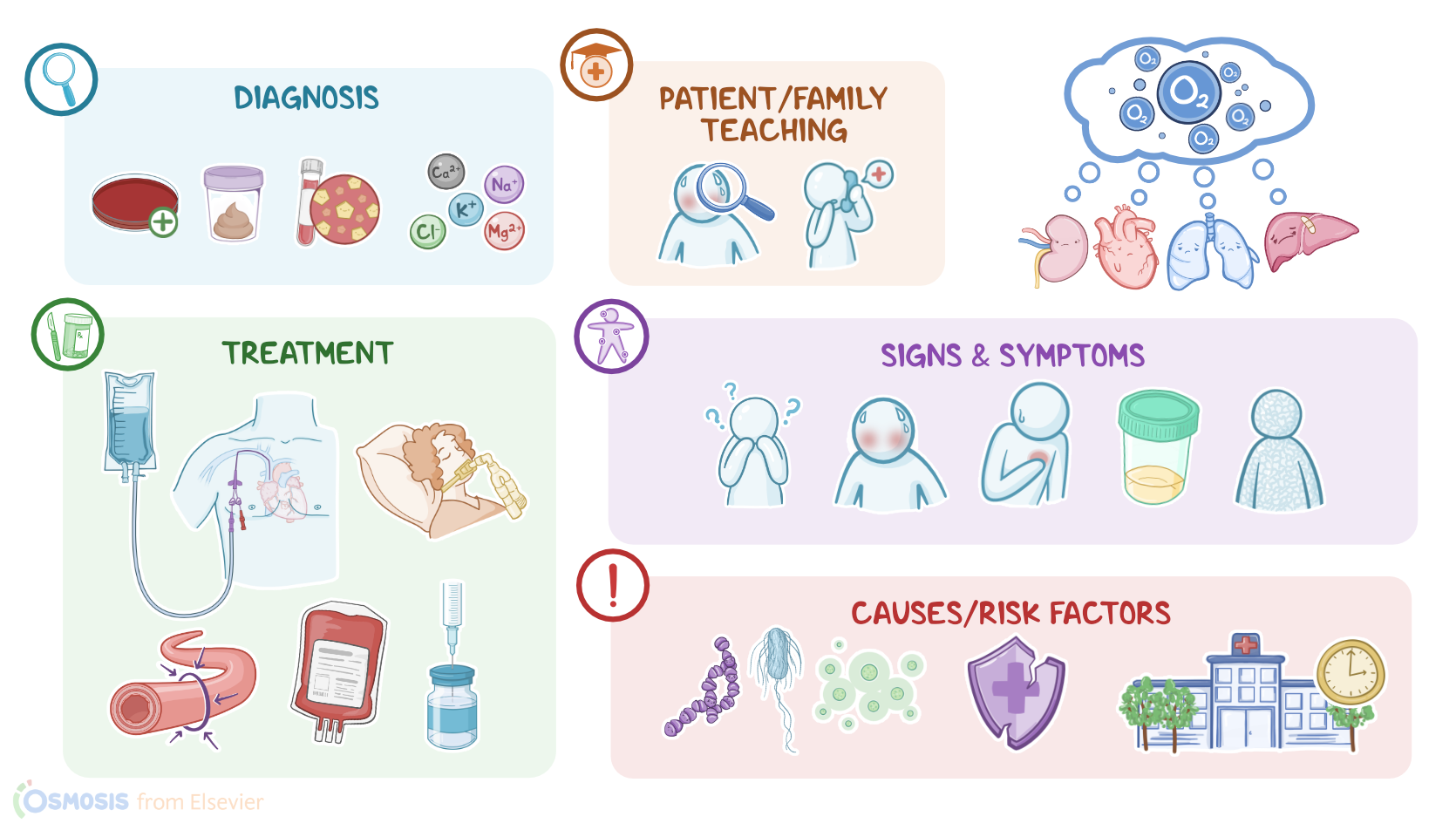
Then, review the signs and symptoms of shock, and stress the importance of seeking medical attention immediately if they experience a fast heart rate; fever or chills; dizziness, fainting, or low blood pressure; shortness of breath; confusion or disorientation; severe pain; or clammy, sweaty skin.Alright, as a quick recap… Septic shock is a type of distributive shock that occurs when the body mounts an excessive inflammatory response because of an infection.
Review14:56–16:40
Clients with compromised immune systems are at increased risk for developing septic shock. This includes children and the elderly, along with clients with an underlying condition like HIV infection, diabetes, chronic kidney or liver disease, or those with chronic glucocorticoid treatment, or those undergoing chemotherapy for cancer treatment.
Other risk factors include prolonged hospital stays, and clients who underwent major surgeries, or have large open wound areas.
Early signs and symptoms of septic shock are fever, warm, flushed skin, tachycardia, respiratory crackles, tachypnea and altered mental status.
Diagnosis of septic shock is confirmed by positive blood cultures, blood tests that show leukocytosis and neutrophilia, increased inflammatory markers, and high lactate levels, among others.
Treatment involves fluid resuscitation and antibiotics, as well as vasopressors like norepinephrine or dopamine; and additional measures include mechanical ventilation, blood replacement therapy and insulin treatment.
Nursing goals of care include recognizing early signs and symptoms of sepsis and intervening to promote hemodynamic stability and adequate organ perfusion.
Client and family education is focused on preventing infection, recognizing signs and symptoms of sepsis, and when to seek medical care.
and adequate organ perfusion. In family, education is focused on preventing infection recognizing signs, and symptoms of sepsis.
And when to seek medical care,
| SHOCK - SEPTIC | ||
| KEY POINTS | NOTES | |
| DEFINITION |
| |
| PHYSIOLOGY |
| |
| CAUSES AND RISK FACTORS |
| |
| PATHOPHYSIOLOGY |
| |
| SIGNS AND SYMPTOMS |
| |
| DIAGNOSIS |
| |
| TREATMENT |
| |
| MANAGEMENT OF CARE |
| |
| PATIENT AND FAMILY TEACHING |
| |
No notes for this video yet
Try adding a note below