Craniopharyngioma · What Is It, Signs & Symptoms, Treatment, and More

Published: Feb 06, 2026
Author: Lahav Constantini, MD
Editor: Alyssa Haag, MD
Editor: Ian Mannarino, MD, MBA
Editor: Kelsey LaFayette, DNP, ARNP, FNP-C
Illustrator: Abbey Richard, MSc
7-day free trial

Go deeper with Osmosis

Osmosis is a learning platform with videos, questions, and AI tools to help you master topics like this.

4.8 · 12,000+ reviews
Watch quick, visual videos
Practice with Qbank-style questions
Use AI to explain, quiz, and review
Study anytime with the mobile app
Start free trial

No credit card · Cancel anytime

What is a craniopharyngioma?

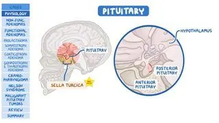
A craniopharyngioma is a rare, benign, and slow-growing central nervous system tumor that may exhibit local aggressive features. The tumor originates from remnants of Rathke pouch, which is an embryogenic structure from which the anterior pituitary gland is formed. The pituitary gland is located within a small cavity in the anterior skull base called sella turcica, which is a bony depression below the hypothalamus. The anatomical structures situated above the sella turcica are termed suprasellar and include the hypothalamus, the optic chiasm, and the circle of Willis. Craniopharyngiomas are frequently located in the sellar and suprasellar region; therefore, the endocrinological, ophthalmological, and vascular systems are at risk for compression by these tumors.  

The incidence of craniopharyngiomas follows a bimodal distribution pattern, peaking in two stages of life: in children aged 5 to 14 years and in adults aged 50 to 74 years. The World Health Organization (WHO) classifies craniopharyngiomas into two distinct tumor types, based on clinical symptoms, demographics, radiologic and histologic findings, and molecular patterns. There is adamantinomatous craniopharyngioma (ACP), which can be found in populations of all ages but is most typical of children, and papillary craniopharyngioma (PCP), which is predominantly found in adults. Most tumors contain both solid and cystic components, and part of the tumor is commonly calcified. 

Learn deeper with Osmosis

Master this topic faster with videos, questions, and AI.

Used by 8M+ healthcare learners.

Start free trial

No credit card · Cancel anytime

What causes a craniopharyngioma?

Craniopharyngiomas are related to distinct gene variants. Adamantinomatous craniopharyngiomas are characterized by activation of the Wnt signaling pathway, most commonly due to pathogenic variants activating CTNNB1. Pathogenic variants of the Wnt signaling pathway lead to excessive formation of β-catenin and its associated oncogenic effects, like cell proliferation and invasion, and tumor formation. Papillary craniopharyngiomas are usually associated with pathogenic variants in the BRAF oncogene, specifically BRAF V600E (i.e., a specific variant in the BRAF gene, where the amino acid valine (V) is replaced by glutamate (E) at position 600). BRAF activates the mitogen-activated protein kinase (MAPK) pathways, which are commonly upregulated in cancers. 

What are the signs and symptoms of craniopharyngioma?

Craniopharyngioma may cause symptoms due to compression of nearby structures. Compression of the optic chiasm where the optic nerves cross, for example, can lead to visual deficits, most commonly bitemporal hemianopsia (i.e., loss of vision in the outer visual fields). Damage or compression of the anterior pituitary can lead to hormonal deficiencies, such as growth hormone deficiency, leading to growth restriction in children. Pressure on the posterior pituitary can lead to vasopressin (i.e., antidiuretic hormone, or ADH) deficiency, formerly known as central diabetes insipidus, which can lead to polyuria and polydipsia. Adults may have a growth hormone deficiency, presenting with weight gain, central obesity, and fatigue; gonadotropin deficiency, manifesting as amenorrhea in genetic females and erectile dysfunction or loss of libido in genetic males; TSH deficiency, causing weight gain, fatigue, cold intolerance, and constipation; and adrenocortical hormone (ACTH) deficiency, presenting with weight loss, arthralgia, dizziness, and hypotension. When the tumor is large, it can obstruct the aqueduct or foramina of Monro and lead to hydrocephalus (i.e., excess buildup of cerebrospinal fluid in the brain ventricles) with a consequent increase in intracranial pressure. Invasion and pressure on the hypothalamus can lead to diencephalic syndrome (i.e., failure to thrive in young children) and obesity. 

Additionally, individuals may present with moderate or severe headaches, sometimes accompanied by nausea, vomiting, and lethargy. Depression and cognitive impairment are also common. 

How is craniopharyngioma diagnosed?

Since craniopharyngiomas are typically slow-growing tumors and the symptoms are related to compression of local structures, diagnosis usually occurs when the associated symptoms begin. The diagnostic process typically includes an assessment of the individual’s medical history and an endocrinological evaluation that includes laboratory tests to evaluate the thyroid and adrenal function (e.g., thyroid-stimulating hormone, ACTH), as well as any hormonal deficiencies that may be present (e.g., follicle-stimulating hormone, luteinizing hormone, estradiol, testosterone). A neuro-ophthalmologic examination assessing visual fields, acuity, and optic disc color and texture may also be performed to evaluate the severity of compression on the optic pathways and identify papilledema. The presence of the mass can be confirmed through magnetic resonance imaging (MRI) or computed tomography (CT scan). CT scan can detect the presence of calcifications, which are a feature of the adult, PCP tumor type. Pathological examination and molecular analysis may confirm the exact diagnosis of the mass. 

The differential diagnoses for other tumors located in the sellar and suprasellar region include pituitary adenoma, germ cell tumors, astrocytomas, and other rare neoplasms and infiltrative disorders. 

How are craniopharyngiomas treated?

Craniopharyngiomas are challenging to treat due to their location near important anatomical structures and frequently recur because they often adhere to nearby structures. Therefore, craniopharyngiomas are typically managed by a multidisciplinary team (MDT) that includes specialists from ophthalmology, endocrinology, neurosurgery, and more, who can collaborate to discern the best course of treatment for the individual.  

Craniopharyngiomas are usually treated with surgical removal, to alleviate the mass-related symptoms. Surgery aims to remove as much of the tumor that can be safely removed. Nonetheless, aggressive surgery, especially for tumors invading the hypothalamus, may lead to significant neuro-endo-vascular-ophthalmological damage. Therefore, a more conservative surgical approach combined with adjuvant therapy is often attempted.   

Since craniopharyngiomas are sensitive to radiotherapy, surgery combined with radiation therapy (using various techniques, such as stereotactic radiosurgery, stereotactic radiotherapy, intensity-modulated radiotherapy, or proton beam radiotherapy) may be the chosen approach. Alternatively, in PCP tumors with the BRAF V600E gene variant, targeted therapy (e.g., vemurafenib and cobimetinib or dabrafenib and trametinib) may be applicable. Histological and molecular assessment of the tumor specimen are necessary to guide treatment. Cystic parts of a tumor can be managed with various operative or nonoperative techniques, such as percutaneous aspiration, intracystic irradiation or chemotherapy, or surgical removal. When hydrocephalus is present, a temporary or permanent shunt can be placed, inserting catheters into the ventricles to drain excess cerebrospinal fluid and relieve pressure.  

All the aforementioned treatment options may result in long-term side effects, especially in children. Damage to the hypothalamus or pituitary stalk, for example, caused by the tumor or exacerbated by treatment, can lead to altered pituitary function that may require long-term treatment with hormone replacement therapy (e.g., levothyroxine in cases of thyroid deficiencies) especially in childhood. Those with vasopressin deficiency may require life-long treatment with vasopressin. Radiotherapy may lead to late visual, endocrine, cognitive, and psychological sequelae, as well as vasculopathies (e.g., Moyamoya disease) and secondary neoplasms (e.g., meningiomas, glioblastomas). The long-term sequelae increase the complexity of craniopharyngioma management and are considered by the MDT when collaborating on the individualized treatment plan 

What are the most important facts to know about craniopharyngiomas?

A craniopharyngioma is a rare, benign, yet locally aggressive tumor of the central nervous system that arises in the sellar or suprasellar areas. It typically develops in two age groups, predominantly children aged 4 to 15 and adults aged 50 to 74. An adamantinomatous craniopharyngioma is a typical tumor that presents in children, while papillary craniopharyngioma predominates in adults. Clinical manifestations include hydrocephalus and compression symptoms, such as visual disturbances, hormonal deficiencies, nausea, vomiting, and headaches. Diagnosis involves endocrinological evaluation, neuro-ophthalmologic examination, as well as neuroimaging and histopathological assessment of the tumor. Treatment options include surgical management, possibly combined with targeted therapy or radiotherapy techniques when the tumor is unable to be completely removed, as well as long-term endocrinological management for any hormonal deficiencies. 

Key Takeaways

Definition 

A craniopharyngioma is a rare, benign, and slow-growing central nervous system tumor that may exhibit local aggressive features. 

Origin and Location 

- Origin: remnants of Rathke pouch 

- Location: sellar and suprasellar region  

     - Risk of compression of endocrinological, ophthalmological, and vascular systems  

Classification 

- Bimodal distribution pattern:  

     - Children 5-14 years  

     - Adults 50-74 years  

- Two tumor types:  

     - Adamantinomatous craniopharyngioma (ACP)  

          - Most typical of children  

     - Papillary craniopharyngioma (PCP) 

          - Most typical of adults  

Causes 

- ACP: Wnt signaling pathway activation (pathogenic variants activating CTNNB1) 

     - Excessive β-catenin → oncogenic effects  

- PCP: pathogenic variant of BRAF (BRAF V600E)   

     - MAPK pathways activation  

Signs and Symptoms 

- Compression of nearby structures:  

     - Optic chiasm → visual deficits  

          - Bitemporal hemianopsia  

     - Anterior pituitary → hormonal deficiencies 

          - Growth hormone deficiency  

          - Gonadotropin deficiency  

          - TSH deficiency  

          - ACTH deficiency  

     - Posterior pituitaryvasopressin (i.e., antidiuretic hormone, or ADH) deficiency 

     - Aqueduct or foramen of Monro (large tumors) → hydrocephalus   

     - Hypothalamus → diencephalic syndrome and obesity  

- Other signs and symptoms:  

     - Moderate / severe headaches  

     - Nausea  

     - Vomiting 

     - Lethargy  

     - Depression  

     - Cognitive impairment  

Diagnosis 

- Usually diagnosed when symptoms from compression of local structures begin  

- Medical history  

- Endocrinological evaluation  

- Neuro-ophthalmologic examination  

- Imaging: MRI, CT scan (mass confirmation)  

     - Calcifications: feature of PCP 

- Pathological examination and molecular analysis 

 - Differential diagnosis: other tumors of sellar and suprasellar region 

     - Pituitary adenoma 

     - Germ cell tumors 

     - Astrocytomas  

Treatment 

- Challenging to treat 

     - Proximity to important anatomical structures 

     - Frequent recurrence (adhere to nearby structures) 

- Multidisciplinary team  

- Surgical removal  

     - Aim: remove as much of the tumor that can be safely removed  

     - Risk of neuro-endo-vascular-ophthalmological damage 

 Conservative surgical approach + adjuvant therapy  

     - Radiation therapy (e.g., stereotactic radiosurgery, stereotactic radiotherapy, intensity-modulated radiotherapy, or proton beam radiotherapy) 

     - Targeted therapy (in PCP tumors with the BRAF V600E gene variant)  

- Cystic parts: percutaneous aspiration, intracystic irradiation or chemotherapy, surgical removal  

- Hydrocephalus: temporary or permanent shunt  

- Side effects of treatments 

     - Damage to hypothalamus or pituitary stalk → altered pituitary function  

          - Hormone replacement therapy might be needed 

     - Radiotherapy → visual, endocrine, cognitive, psychological sequelae, vasculopathies, secondary neoplasms  

Students say Osmosis is 100% worth it

Because Osmosis saves them time. Lowers stress. And actually helps them remember when it counts.

I used Osmosis to prepare for my first medical school licensing exam! Super helpful and interactive for people who may not do great with just pages of text info!

Cecilia Ruiz

Cecilia Ruiz

MD student

Sayan Misra

I have used Osmosis for about four years. Best thing I have ever used for my medical studies.

Sayan Misra

Sayan Misra

Med student

Osmosis videos are superior because they define simple concepts, tell a story with a clear progression, and provide context.

Jay Pate

Jay Pate

Dental student

References


Daroff RB, Jankovic J, Mazziotta JC, Scott Loren Pomeroy, Bradley WG. Bradley’s neurology in clinical practice. Elsevier; 2016. 


Gabay S, Merchant TE, Boop FA, Roth J, Constantini S. Shifting strategies in the treatment of pediatric craniopharyngioma. Curr Oncol Rep. 2023;25(12):1497-1513. doi:10.1007/s11912-023-01471-9  


Garrè ML, Cama A. Craniopharyngioma: Modern concepts in pathogenesis and treatment. Curr Opin Pediatr. 2007;19(4):471-479. doi:10.1097/MOP.0b013e3282495a22 


Khan RB, Merchant TE, Boop FA, et al. Headaches in children with craniopharyngioma. J Child Neurol. 2013;28(12):1622-1625. doi:10.1177/0883073812464817 


Jane JA Jr, Laws ER. Craniopharyngioma. Pituitary. 2006;9(4):323-326. doi:10.1007/s11102-006-0413-8  


Louis DN, Perry A, Wesseling P, et al. The 2021 WHO Classification of tumors of the central nervous system: A summary. Neuro Oncol. 2021;23(8):1231-1251. doi:10.1093/neuonc/noab106 


Müller HL, Merchant TE, Warmuth-Metz M, Martinez-Barbera JP, Puget S. Craniopharyngioma. Nat Rev Dis Primers. 2019;5(1):75. Published 2019 Nov 7. doi:10.1038/s41572-019-0125-9