Hemolytic disease of the fetus and newborn: Nursing
Introduction0:00–0:18
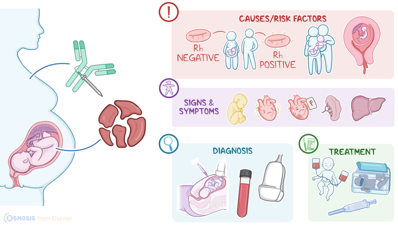
Hemolytic disease of the fetus and newborn, also called erythroblastosis fetalis, is an immune-mediated condition in which the red blood cells of the fetus or newborn are hemolyzed, or destroyed by the mother’s antibodies.
Physiology0:18–2:25
Let’s start by looking at the physiology of blood groups. The most common blood group classification is based on the ABO and the Rh system.
These systems are based on the presence or absence of certain glycoproteins on the surface of red blood cells. The ABO system relies on the presence or absence of type A and B glycoproteins on RBCs.
So people with the A blood type have type A glycoproteins; people with B blood type have type B glycoproteins; people with type AB, intuitively, have both; and people with the O blood type have neither type of glycoproteins on their RBCs.
The immune system produces antibodies against the glycoproteins that are missing from the surface of the individual’s erythrocyte.
That’s why people with type A blood have antibodies to type B blood, and those with type B blood have antibodies to type A blood.
People with blood type AB don’t have antibodies to type A or type B blood; whereas people with type O blood have antibodies to both A and B glycoproteins.Next, the Rh classification looks at whether or not the Rhesus factor, or “Rh” factor for short, is present on RBCs.
If this factor is present on their red blood cells, that makes the blood type Rh positive; whereas if it’s absent, the blood type is Rh negative.
People that are Rh positive can receive both Rh negative or Rh positive blood since they do not have antibodies against the Rh glycoprotein.
But if an Rh negative person receives Rh positive blood, they could develop a hemolytic transfusion reaction. Having said that, Rh antibodies require prior exposure to Rh positive RBCs.
So, someone who doesn’t have Rh antibodies, won’t have a transfusion reaction the first time they’re exposed to Rh positive blood.
However, they develop anti-Rh antibodies, so if they’re exposed to Rh positive blood a second time, that will cause a transfusion reaction.
Causes & risk factors2:25–3:21
Now, the hemolytic disease of the newborn is mediated by the immune system and can occur when fetal erythrocytes, which express an RBC antigen not expressed on maternal RBCs, breach into maternal circulation, usually when placenta separates at birth.
This most commonly occurs due to Rh incompatibility; less commonly, it can be due to ABO incompatibility between the mother and the fetus.
Risk factors include the mother being Rh negative while the conceiving partner is Rh positive; second or higher pregnancy; previous pregnancies with hemolytic disease; previous miscarriages or pregnancy complications; Caucasian race; pregnant individuals that have not been tested or received prevention with Rh immunoglobulin; and invasive medical procedures like amniocentesis or chorionic villous sampling.Pathology-wise, if the baby’s RBCs have any the ABO or Rh glycoproteins that the mother’s immune system hasn’t previously encountered, then these glycoproteins can act as antigens, causing the mother’s immune system to produce antibodies against them.
Pathophysiology3:21–4:49
Now, in a first pregnancy, this does not affect the fetus. However, in a subsequent pregnancy, if the fetus is again positive for the same glycoprotein, like, for example, if the fetus is Rh positive, then the mother’s immune system mounts a fast and specific attack against the fetal RBCs, causing hemolysis.
Now, hemolytic disease of the fetus and newborn has significant consequences during pregnancy and after delivery. During pregnancy, hemolysis can result in mild fetal anemia, which can cause fetal tachycardia.
In case of severe anemia, fetal heart failure can result, causing a buildup of fluids in various organs and tissues, including pericardial effusion, pleural effusion, abdominal ascites, as well as liver and spleen enlargement.
This is a condition called hydrops fetalis, and it can be fatal. Moreover, as bilirubin starts to accumulate secondary to hemoglobin breakdown, hyperbilirubinemia develops too, causing jaundice.
In utero, jaundice can be seen as a yellow discoloration of the amniotic fluid or umbilical cord. After birth, bilirubin buildup can damage the brain, a condition called kernicterus.The presentation after birth varies depending on the severity of the hemolysis.
Clinical manifestations4:49–5:26
With mild anemia, the infant’s vital signs are usually normal, and jaundice develops within 24 hours after birth. Moderate anemia can cause pallor, tachycardia, and jaundice, also within the first 24 hours of life.
If the anemia is severe and the baby develops hydrops fetalis, there could be edema or swelling of the entire body, extreme pallor, and significant difficulty in breathing due to heart failure.
On palpation, there could also be an enlarged liver and spleen. Diagnosis of hemolytic disease of the fetus and newborn starts with a thorough history and physical examination.
Diagnosis5:26–6:41
During pregnancy, diagnosis can begin with blood typing for ABO and Rh status of the parents early in the first trimester.
Fetal blood type and the presence of antibodies against fetal RBCs can also be determined, typically through amniocentesis.
This procedure can also be used to measure bilirubin levels in the amniotic fluid. A prenatal ultrasound can also help identify signs of hydrops fetalis or organ enlargement.
After birth, diagnosis of hemolytic disease is confirmed with either a positive direct or indirect antiglobulin test, like the Coombs test, which will confirm the presence of maternal RBC antibodies in the infant’s serum.
Additionally, the baby’s blood can be tested for type; a CBC may show low hemoglobin and RBC levels, and high reticulocyte count.
Management involves prevention, and prenatal and postnatal treatment. Prevention starts at the first prenatal visit to determine maternal Rh status and sensitization.
Treatment6:41–8:46
If an Rh negative mother has not been sensitized, Rh immunoglobulin is given to reduce the risk of developing antibodies against the fetus’ potentially Rh positive RBCs.
After birth, the newborn should have their blood group and Rh status determined, and if they’re Rh positive, the mother should receive a second dose of Rh immunoglobulin within 72 hours after delivery.
Prenatal treatment management depends on the severity of the condition. Mild cases can be managed by watchful waiting and cord blood sampling to test for fetal anemia.
Some cases may require intrauterine transfusions. Fetal ultrasonography can be used to check for evidence of generalized fetal edema, hepatosplenomegaly, or hydrops.
In severe cases of fetal anemia, labor and delivery induction or cesarean delivery might be necessary, along with delayed cord clamping to increase the infant’s hemoglobin level.After birth, most cases of hemolytic disease are managed by treating hyperbilirubinemia with phototherapy.
Phototherapy uses light to transform bilirubin into a water-soluble isomer that can then be excreted by the infant’s kidneys and stool without the need to be processed in the liver.
If phototherapy fails or in cases of severe disease, exchange transfusions might be needed. This involves replacing infant RBCs with antigen-negative RBCs, preventing further hemolysis.
Another approach can be to give intravenous immunoglobulin, which contains antibodies to help the baby’s immune system and reduce hemolysis.
Finally, supportive measures might be necessary, including packed RBC transfusions, IV fluids, and supplemental oxygen or mechanical ventilation.Okay, let’s talk about the care you’ll be giving to an infant with hemolytic disease of the newborn.
Management and care8:46–9:46
Your primary goal will be to manage complications. First, assess the infant’s vital signs, observe their energy level and feeding ability, assess their skin, eyes, and mucous membranes, and draw lab work as ordered.
Report to the healthcare provider for tachycardia or tachypnea; pale, cyanotic or jaundiced skin or mucous membranes; lethargy, poor feeding; or abnormal lab work such as a low RBCs, high reticulocyte count, or high bilirubin level.
Provide supplemental oxygen for respiratory support and place an IV to begin the prescribed fluids for hydration and promotion of urine and stool output.
Also administer the prescribed IV immunoglobulin to help reduce hemolysis. Then, begin phototherapy to reduce bilirubin levels, or prepare for a blood exchange transfusion if severe anemia or hyperbilirubinemia is present.All right, let’s move on to client and family teaching.
General client and family teaching9:46–10:43
Begin by explaining to the caregivers that hemolytic disease of the fetus and newborn is caused when the baby's red blood cells break down because of an incompatibility between the mother’s blood type and the baby’s blood type, causing anemia and high levels of bilirubin.Next, explain that when bilirubin levels are too high, their baby needs to be placed under special lights called phototherapy that will help the body get rid of the extra bilirubin and prevent problems like brain damage.
Finally, when the infant is ready to be discharged home, remind them to keep all follow-up appointments. Instruct them to notify their provider if there’s new or worsening yellowing of baby’s skin or if they seem less active or less interested in feeding.
Stress the importance of contacting or seeking emergency medical care if their baby develops a fever or has trouble breathing.All right, as a quick recap… Hemolytic disease of the fetus and newborn is a condition in which red blood cells of the fetus or newborn are destroyed by antibodies from the mother.
Review10:43–11:56
This process is mediated by the immune system, and most commonly occurs when a mother with a Rh negative blood type has been sensitized to and has produced antibodies against an antigen present in Rh positive blood type.
When these antibodies cross the placenta into the bloodstream of a Rh positive fetus, the fetal red blood cells are destroyed, often resulting in anemia and hyperbilirubinemia.
Diagnosis is made during pregnancy with blood typing of the parents and possible amniocentesis, and after birth with a blood test showing the presence of maternal antibodies in the infant’s blood.
Treatment focuses on prevention through immunoglobulin administration, prenatal monitoring, and phototherapy, exchange transfusions, and immunoglobulin for the newborn after birth.
The priority nursing goal is to manage complications, and client and family education centers on explaining the condition, discussing treatments that may be needed, and when to contact their healthcare provider.
| HEMOLYTIC DISEASE OF THE FETUS AND NEWBORN | ||
| KEY POINTS | NOTES | |
| DEFINITION |
| |
| PHYSIOLOGY |
| |
| CAUSES AND RISK FACTORS |
| |
| PATHOPHYSIOLOGY |
| |
| SIGNS AND SYMPTOMS |
| |
| DIAGNOSIS |
| |
| TREATMENT |
| |
| MANAGEMENT OF CARE |
| |
| PATIENT AND FAMILY TEACHING |
| |
No notes for this video yet
Try adding a note below