Definitions & Key takeaways

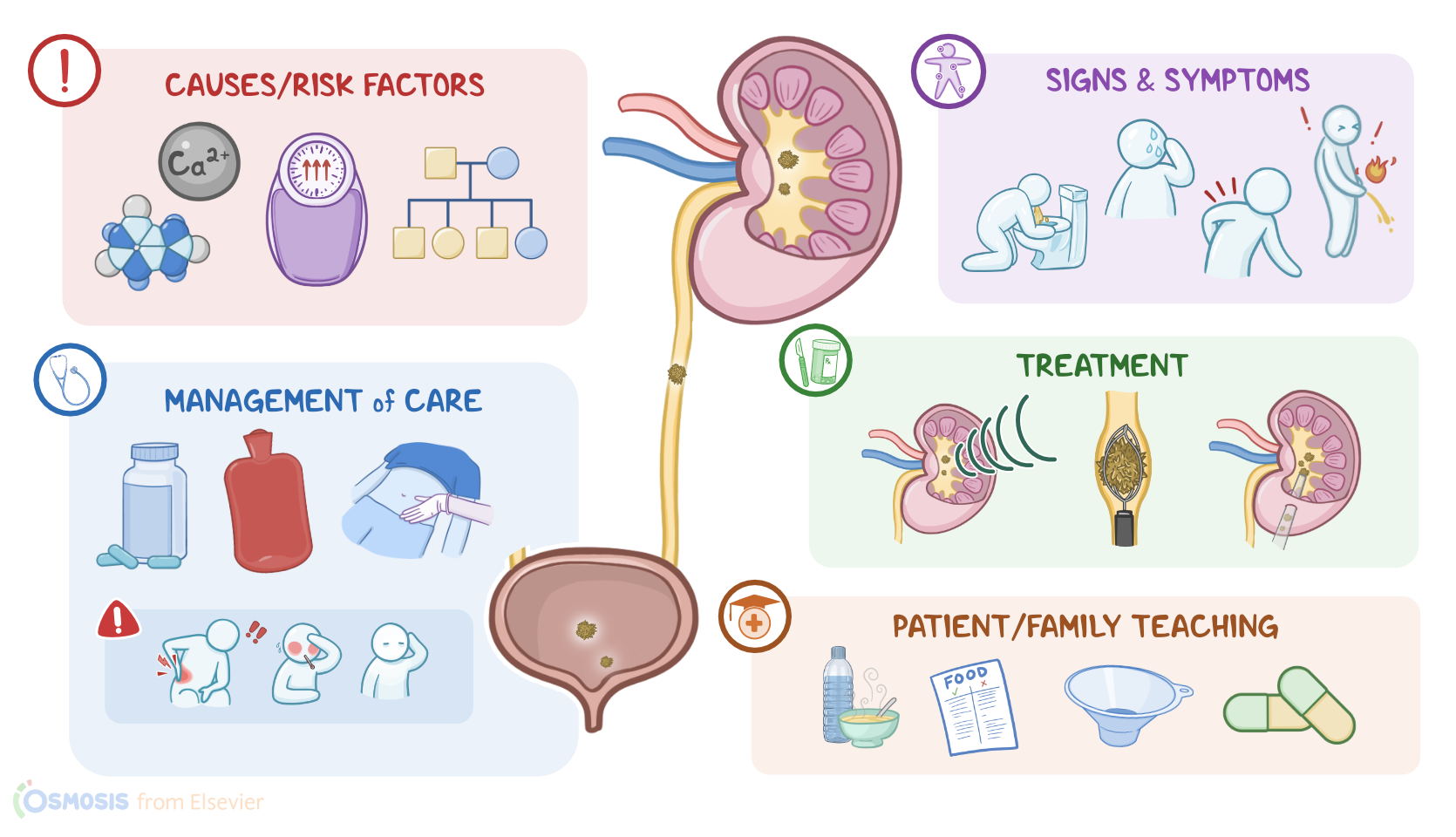
Nephrolithiasis and urolithiasis refer to stones, also called calculi, in the kidney and urinary tract, respectively. They form when solutes in the urine precipitate out and crystallize. Depending on the type of precipitated solute forming the stone, there are five main types of stones: calcium oxalate, calcium phosphate, struvite, uric acid, and cystine stones. Renal and urinary stones can cause urinary tract obstruction, infection, and even hydronephrosis, which can lead to permanent kidney damage.

Even though people with renal and urinary stones may be asymptomatic, common symptoms include renal colic and flank pain. The pain is often sharp, sudden, and severe enough that it may trigger nausea, vomiting, pallor, and sweating. There may also be hematuria, dysuria, urinary urgency and frequency, and signs of infection.

The diagnosis involves a history and physical assessment, followed by lab exams such as urinalysis and blood studies, as well as imaging like CT and ultrasound. Treatment includes medications to treat pain and surgery in some severe cases. There are also other procedures like extracorporeal shock wave lithotripsy (ESWL), which uses high-energy sound waves to break these stones into smaller fragments able to be passed in the urine. Increased fluid intake and dietary changes may be recommended to prevent stone formation in the future.

Chapters:

Introduction0:00–0:29

Nephrolithiasis and urolithiasis refer to the presence of stones, also known as calculi, in the kidney and the urinary tract, respectively.
Stones form when solutes in the urine precipitate out and crystallize. Depending on which solute precipitates to form the stone, there are five main types of stones: calcium oxalate, calcium phosphate, struvite, uric acid, and cystine stones.Now, let’s quickly review some anatomy and physiology of the urinary tract, which is made of the kidneys, ureter, bladder, and urethra.

Physiology0:29–1:43

The kidneys are in charge of producing urine, which is made up of water and solutes. The process starts with glomerular filtration in the renal corpuscle, which is made up of the glomerulus and the Bowman’s capsule.
As blood flows into the glomerulus, water, electrolytes like sodium, and other small particles in the blood like creatinine, urea nitrogen, and glucose, pass through the endothelial lining of the capillaries and into Bowman's space.
Large proteins and red blood cells however, aren’t filtered. Now, the next steps, which include tubular reabsorption and secretion, take place in the renal tubules, which are surrounded by the peritubular capillaries.
Here, water and solutes get passed back and forth between the filtrate in the lumen of the renal tubule and the blood in the peritubular capillaries.
Finally, urine is drained into the renal pelvis, which then narrows to form the ureter, and transports urine to the bladder.
Ultimately, urine exits the bladder through the urethra.Now, nephrolithiasis and urolithiasis are caused by the increased concentration of certain solutes in the urine.

Causes & risk factors1:43–5:06

This can happen if there’s an increase in the solute, which could be calcium with oxalate or phosphate, leading to the formation of calcium stones; magnesium, ammonia, and phosphate, leading to struvite stones, as well as uric acid forming uric acid stones and cystine, forming cystine stones.
Alternatively, there could be a decrease in the solvent, or water in the urine. That being said, the exact cause why these stones form is usually unknown, but they have been associated with several risk factors.
Common risk factors for all types of kidney stones include a family history of nephrolithiasis, prolonged immobility, obesity, and dehydration, which is more likely to occur in warm climates or due to low fluid intake.
Now, for calcium stones, risk factors mainly include hypercalcemia and hypercalciuria. This can be a complication of hyperparathyroidism, because parathyroid hormone increases calcium release from the bones and calcium reabsorption from the intestines, while decreasing calcium excretion from the kidneys.
Hypocitraturia is another cause, where less citrate is eliminated in the urine. This is because normally, citrate prevents kidney stone formation by inhibiting crystal growth and aggregation.
Additionally, there are medications that can increase the risk of developing calcium stones, like loop diuretics, acetazolamide, theophylline and glucocorticoids.
Other risk factors include fat malabsorption, ingestion of ethylene glycol or antifreeze, and high levels of vitamin C, all of which raise oxalate levels in the blood.
Finally, a diet heavy in oxalate-rich foods like rhubarb, spinach, chocolate, nuts, and beer increases the risk of calcium stones.
For uric acid stones, risk factors include acidic urine, which can be caused by acidosis, meaning a low blood pH. This can happen with chronic diarrhea, where large amounts of bicarbonate are lost in the stool; or with poorly controlled diabetes, where the body can’t use glucose and instead breaks down fat for fuel, creating acidic ketones as a by-product.
Another important risk factor is hyperuricemia, or high levels of uric acid in the blood, which might be due to conditions like gout.
There are also medications that promote the development of uric acid stones, like thiazide diuretics, salicylates, probenecid and allopurinol.
Lastly, since uric acid is a breakdown product of purine, consuming lots of purines, which can be found in shellfish, anchovies, red meat or organ meat, increases the risk of uric acid stones.
Next are cystine stones, which occur in the setting of cystinuria, a genetic condition where cystine cannot get reabsorbed from the renal tubules, so instead gets excreted in the urine.
Finally, for struvite stones, the main risk factor is a urinary tract infection with bacteria that produce urease, like Proteus and Klebsiella.
These bacteria increase urine pH, making it a favorable environment for magnesium ammonium phosphate to precipitate.Alright so, pathology of kidney stones begins with urinary supersaturation of certain solutes, which results in precipitation and formation of crystals.

Pathology5:06–6:04

Those crystals then act as a nidus, or place where more solutes can deposit, and, over time, this forms a kidney stone. Now, the stone can become stuck and obstruct the urinary tract, which happens most commonly in ureteropelvic junction or in the ureter.
When this happens, the ureter becomes dilated and stretched. At the same time, the urothelial lining is damaged, which causes bleeding and consequent hematuria.
Also, if the obstruction persists, urine won’t flow, which is called urinary stasis, making the urinary tract prone to infection.
Sometimes, kidney stones can completely obstruct the ureter and this way the urine builds up behind the obstruction, causing hydronephrosis, or dilation of the renal pelvis and calyces, which can cause permanent kidney damage.
Now, the clinical manifestations of lithiasis depend on the location of the stones. Sometimes, clients with kidney stones can be asymptomatic.

Clinical manifestations6:04–6:53

Other times they can present with renal colic, which is when there’s acute lumbar pain due to urinary obstruction and renal distention.
When the stone lodges in the ureter, it can cause unilateral flank pain that can radiate to the testicle or labia on the affected side.
This pain is typically sharp, of sudden onset, and so intense that it can cause nausea, vomiting, pallor, and sweating. In addition, there can also be frank hematuria.
Finally, if the stone gets lodged in the distal ureter, there might be clinical manifestations of urinary tract infection, like dysuria, urinary urgency, urinary frequency, and fever.Diagnosis of nephrolithiasis and urolithiasis involves history and physical assessment, followed by urinalysis and blood studies.

Diagnosis6:53–7:48

In urinalysis it’s important to look for the presence of hematuria, white blood cells, crystals, and bacteria, as well as checking the urine pH.
A high urine pH is indicative of a struvite stone; whereas a low urine pH typically suggests a uric acid stone. Blood studies can show leukocytosis when there’s infection.
Electrolytes, creatinine, BUN, calcium, uric acid, phosphorus, and parathyroid hormone levels might also help determine the type of stones.
High serum or urinary calcium levels, as well as low potassium and bicarbonate levels usually indicate calcium stones. Finally, an ultrasound or a non-contrast CT scan can help directly visualize the stones.The treatment of nephrolithiasis and urolithiasis depends on the clinical manifestations and the location, size, and composition of the stones.
For individuals with acute renal colic, pain is managed with NSAIDs or opioids. If symptoms are controlled and the stones are small, then medical expulsive therapy, or MET, might be used to help pass the stone.

Treatment7:48–9:34

This typically requires an alpha-adrenergic blocker like tamsulosin or a calcium channel blocker, like nifedipine to relax the smooth muscle in the ureter.
Now, when MET doesn’t work, symptoms are severe and persistent, or stones are larger, then a more aggressive approach is usually taken.
One option is extracorporeal shock-wave lithotripsy, or ESWL, which uses high-energy sound waves that produce shock waves to break kidney stones into smaller fragments which then can be passed in the urine.
Then there’s flexible ureteroscopy, or URS, which is done with an endoscope that’s passed through the urethra, bladder, and finally into the ureter where it’s used to remove the stone.
Another option is percutaneous nephrolithotomy, or PCNL, a minimally-invasive procedure where a small incision is made in the flank and then a flexible nephroscope is inserted and the stone is removed through the working canal of the nephroscope.
Now, when there’s persistent pain, infection or obstruction, surgery is indicated, and depending on the location of the stones, the chosen procedure can be a nephrolithotomy, pyelolithotomy, ureterolithotomy, or cystolithotomy.
Finally, an increased fluid intake and dietary changes are recommended to prevent stone formation in the future.Alright, now let’s look at the nursing care you’ll provide for a client with a renal or urinary calculi.
Your priority nursing goals are to manage pain, promote stone expulsion, and monitor for complications during treatment.

Management and care9:34–11:29

Begin by assessing your client's pain level, noting the location and severity, and check for nausea or vomiting. Administer the prescribed analgesics to decrease pain, and provide comfort measures by guiding your client through relaxation techniques and applying warm compresses to the affected side.
Report to the healthcare provider immediately if your client’s pain is unrelieved by medications or if their pain worsens.
Prepare your client for ESWL or surgical intervention, as indicated. Then, promote stone expulsion by administering the alpha-adrenergic blocker medication as prescribed.
Also, initiate IV fluids and assist them with frequent ambulation. Make sure to strain their urine so when a stone is expelled it can be sent to the lab for analysis.
Next, monitor your client for complications of renal or urinary calculi. Assess their skin color and temperature, monitor their fluid intake and output, check bladder distention, and perform a bladder scan to assess for urinary retention.
Report to the healthcare provider right away if your client becomes pale, diaphoretic, or has decreased urinary output or bladder distention, because these signs could indicate an obstruction.
Then prepare your client for surgical intervention, as indicated. Also, assess your client’s vital signs, urine color and odor, and review their laboratory test results to monitor for infection.
Immediately report signs of infection, such as a fever, dysuria, foul smelling urine, or elevated WBCs. Obtain cultures and administer antibiotics as prescribed.Okay, let's move on to client and family education.
First, explain to your client that renal and urinary calculi are stones that form in the kidney or urinary tract that can cause pain and block the flow of urine.
Discuss the type of stone that caused their pain, if identified, and allow time for them to ask questions. Next, encourage your client to take their prescribed medications exactly as directed, and emphasize the importance of keeping follow-up appointments for continued monitoring.

General client and family teaching11:29–12:37

Then, teach your client ways to prevent stone formation, such as drinking enough fluids to keep the urine pale yellow. Provide resources to assist your client with healthy food choices and teach them about which foods to avoid to prevent future stone formation.
If your client is discharged home before the stone is expelled, show them how to use a strainer to strain their urine to catch the stone.
Finally, advise your client to notify the healthcare provider immediately if they experience severe pain unrelieved by pain medication, decreased urinary output, bladder distention, blood in the urine, painful urination, or fever.
Alright, as a quick recap… Nephrolithiasis and urolithiasis refer to stones, also called calculi, in the kidney and urinary tract, respectively.
There are five main types of stones: calcium oxalate, calcium phosphate, struvite, uric acid and cystine stones. Stones are caused by increased concentration of certain solutes in the urine, either due an increase in the solute or a decrease in the solvent.

Review12:37–14:26

The exact cause of stone formation is unknown, but a family history and dehydration are general risk factors, in addition to specific risk factors that vary depending on the type of stone.
Stones can cause obstruction, hematuria, infection, and even hydronephrosis which can lead to permanent kidney damage. Now, a client with a kidney stone can be asymptomatic.
However, typical manifestations include renal colic and flank pain. The pain is often sharp, sudden and severe enough that it may cause nausea, vomiting, pallor, and sweating.
Other manifestations include hematuria, dysuria, urgency, urinary frequency and infection. Diagnosis begins with a history and physical assessment, followed by urinalysis and blood studies, as well as imaging like CT and ultrasound.
Treatment includes medications to treat pain and medical expulsive therapy. In some cases procedures like ESWL, URS, or PCNL may be necessary, or even surgical stone removal.
Finally, increased fluid intake and dietary changes are recommended to prevent stone formation in the future. Nursing management focuses on providing pain management, promoting stone expulsion, and monitoring for complications.
Client and family education emphasizes prevention of future stone formation and when to seek medical treatment. pcnl, may be necessary.
Or even surgical stone removal, finally increased fluid intake, and dietary changes are recommended to prevent Stone formation in the future.
Nursing management. Focuses on, providing pain management, promoting Stone expulsion and monitoring for complications client and family.
Education emphasizes prevention of future Stone formation. And when to seek medical treatment,