Bladder exstrophy: Nursing

Last updated: September 14, 2022

Bladder exstrophy: Nursing

Pediatrics

Pediatrics

Pediculosis and scabies: Nursing
Atopic dermatitis: Nursing process (ADPIE)
Childhood nutrition and obesity: Information for patients and families (The Primary School)
Childhood oral health: Information for patients and families (The Primary School)
Chickenpox (Varicella): Nursing process (ADPIE)
Appendicitis: Nursing process (ADPIE)
Fractures: Nursing process (ADPIE)
Hirschsprung disease: Nursing
Rheumatic heart disease: Nursing process (ADPIE)
Carbon monoxide poisoning: Nursing process (ADPIE)
Conjunctivitis: Nursing process (ADPIE)
Reye syndrome: Nursing process (ADPIE)
Congenital heart defects - Acyanotic: Nursing
Congenital heart defects - Cyanotic: Nursing
Kawasaki disease: Nursing
Strabismus: Nursing
Otitis media: Nursing
Eye injury: Nursing process (ADPIE)
Tonsillitis: Nursing process (ADPIE)
Cleft lip and palate: Nursing
Esophageal atresia and tracheoesophageal fistula: Nursing
Omphalocele and gastroschisis: Nursing
Poisoning: Nursing process (ADPIE)
Pyloric stenosis: Nursing process (ADPIE)
Bladder exstrophy: Nursing
Cryptorchidism: Nursing
Enuresis: Nursing
Hypospadias and epispadias: Nursing
Hemolytic uremic syndrome: Nursing
Sickle cell disease: Nursing process (ADPIE)
Erythema infectiosum (Fifth disease): Nursing
Fever: Nursing
Infectious mononucleosis: Nursing
Pertussis: Nursing
Poliomyelitis: Nursing
Rocky Mountain spotted fever (RMSF): Nursing
Rubeola (Measles): Nursing
Zika virus: Nursing
Lyme disease: Nursing process (ADPIE)
Child maltreatment: Nursing
Crisis intervention: Nursing
Therapeutic communication: Nursing
Autism spectrum disorder (ASD): Nursing
Failure to thrive (FTT): Nursing
Growth and development theories: Nursing
Growth and development - Adolescent: Nursing
Growth and development - Infant: Nursing
Growth and development - Preschool-age: Nursing
Growth and development - School-age: Nursing
Growth and development - Toddler: Nursing
Sudden infant death syndrome (SIDS): Nursing
Muscular dystrophies - Duchenne and Becker: Nursing
Clubfoot: Nursing
Scoliosis: Nursing
Developmental dysplasia of the hip: Nursing
Juvenile idiopathic arthritis: Nursing
Cerebral palsy: Nursing
Hydrocephalus: Nursing process (ADPIE)
Influenza: Nursing
Asthma: Nursing process (ADPIE)
Bronchiolitis and respiratory syncytial virus (RSV): Nursing process (ADPIE)
Epiglottitis: Nursing process (ADPIE)
Foreign body aspiration and upper airway obstruction: Nursing process (ADPIE)
Laryngotracheobronchitis (LTB) and croup: Nursing process (ADPIE)
Smoke inhalation injury: Nursing process (ADPIE)
Cystic fibrosis: Nursing
Head injury: Nursing
Pediatric psychosocial needs during illness and hospitalization: Nursing
Diarrhea: Nursing
Metabolic alkalosis
Metabolic acidosis
Shock - Hypovolemic: Nursing
Shock - Septic: Nursing
Respiratory acidosis
Acid-base map and compensatory mechanisms
Seizure disorder: Nursing process (ADPIE)
Cutaneous fungal infections: Nursing
Precocious puberty: Nursing
Tumor lysis syndrome (TLS): Nursing Process (ADPIE)
Lymphoma - Hodgkin and non-Hodgkin: Nursing
Leukemia: Nursing process (ADPIE)
Hemophilia: Nursing process (ADPIE)
Anemia - Iron-deficiency: Nursing
Biology of cancer: Nursing
Increased intracranial pressure (ICP): Nursing
Spinal cord injury (SCI): Nursing
Neurological assessment - Neonate: Nursing
Neural tube defects: Nursing
Meningitis: Nursing process (ADPIE)
Amblyopia: Nursing
Hearing impairment and otosclerosis: Nursing
Case study - Pediatric appendicitis: Nursing
Case study - Pediatric anaphylaxis: Nursing
Case study - Pediatric urinary tract infection: Nursing
Case study - Pediatric diabetes mellitus type 1: Nursing
Case study - Child maltreatment: Nursing
Case study - Autism spectrum disorder: Nursing
Case study - Sickle cell anemia: Nursing

Notes

BLADDER EXSTROPHY

KEY POINTS
NOTES
DEFINITION
  • Congenital malformation where bladder protrudes outside the abdomen

PHYSIOLOGY
  • Bladder development
    • Begins during 4th week
    • Three germ layers
      • Ectoderm
      • Mesoderm
      • Endoderm
    • Urogenital sinus forms primitive bladder

CAUSES AND RISK FACTORS
  • Causes
    • Midline closure defect during development
      • Unknown
  • Risk factors
    • Family history
    • White race
    • Assigned male at birth
    • Assisted reproductive technology

PATHOPHYSIOLOGY
  • Normal midline closure disrupted
  • Complications
    • Urinary incontinence 
    • Risk of urinary tract infections
    • Sexual dysfunction
    • Risk of bladder cancer

SIGNS AND SYMPTOMS
  • Inside of bladder on outside of abdomen
  • Urine dripping from open bladder
  • Low-set umbilicus
  • Diastasis of symphysis pubis
  • Other genital malformations

DIAGNOSIS
  • Prenatal ultrasound
  • Observation

TREATMENT
  • Surgery

MANAGEMENT OF CARE
  • Goals of care
    • Protect bladder
    • Monitor for complications
  • Cover bladder with non-adherent dressing
  • Place infant in isolette
  • Monitor temperature
  • Administer medications as prescribed
  • Keep in supine position 
  • Change diapers immediately upon soiling
  • Use latex-free gloves and equipment 
  • Assess pain
  • Decrease environmental stimuli
  • Cluster care
  • Monitor intake and output
    • Notify HCP
      • Urinary output less than 0.5-2 mg/kg/hour
  • Review laboratory results 
  • Monitor surgical site and urine
    • Notify HCP
      • Signs of infection

PATIENT AND FAMILY TEACHING
  • Explain condition, plan of care, and how to safely administer medications
  • Teach caregivers how to change diapers and keep surgical site clean
  • Notify HCP
    • Signs of infections
    • Problems with urination

Transcript

Watch video only

Bladder exstrophy is a congenital malformation where the bladder protrudes outside of the abdomen due to an abdominal wall defect.

First, let’s quickly review the embryological development of the bladder. The renal system, including the bladder, starts developing during week 4 of intrauterine life. At that point, the embryo is made up of three primitive germ layers: the ectoderm, the mesoderm and the endoderm. Towards the end of week 4 of development, the endoderm in the hindgut, which is the last portion of the primitive digestive tract, begins to expand forming the cloaca. The cloaca is a temporary structure connecting the urinary, digestive, and reproductive ducts.

By week 7, a wall of tissue called the urorectal septum forms in the cloaca and splits it into a posterior anorectal canal for the digestive tract, and an anterior urogenital sinus for genital and urinary structures. And the top portion of the urogenital sinus stretches out to form the primitive bladder. Ultimately, the cloacal membrane opens to the outside of the body for these structures to have openings. Now, at around the same time in development, the muscles and connective tissue of the lower anterior abdominal wall also begin to form.

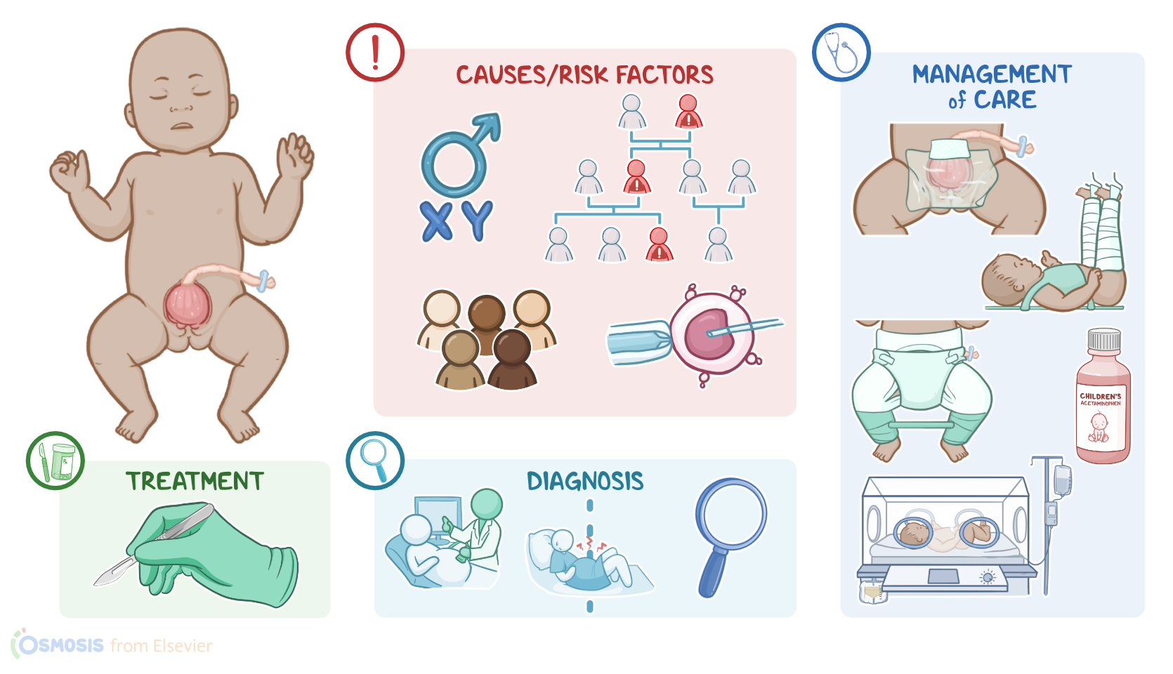
Alright now, bladder exstrophy occurs when there is a midline closure defect during fetal development. Although the exact cause remains unknown, several risk factors have been identified. These include having a family history of bladder exstrophy, being white, or being assigned male sex at birth. This condition is also more common in infants born through assisted reproductive technology, such as in vitro fertilization, or IVF.

Now, when it comes to pathology, the most widely accepted theory for how bladder exstrophy occurs is that the cloacal membrane grows too much, and pushes towards the front of the embryo like a wedge. This prevents the normal midline closure of the abdominal wall. Subsequently, the cloacal membrane ruptures, which renders lower abdominal structures, like the bladder, open and exposed outside of the lower abdominal wall. This also affects the development of other lower abdominal structures, including the symphysis pubis, genitalia and anus.

Okay, now bladder exstrophy can lead to several complications, including urinary incontinence and an increased risk of urinary tract infections. It can also lead to sexual dysfunction and an increased risk of bladder cancer.

Now, individuals with bladder exstrophy present with the inside of the bladder being outside of the abdomen. The bladder is also usually flat instead of its typical rounded shape. Since the bladder is open, urine often drips from the open bladder rather than through the urethra. Individuals with bladder exstrophy can also have a low-set umbilicus; diastasis of the symphysis pubis, meaning this joint is often wider than normal; and an anteriorly displaced anus, where the anal opening is placed more towards the front.

Other genital malformations are also commonly present. In assigned males at birth, these can include a shortened penis and epispadias, which is when the urethra opens on the dorsal side, or on top of the penis. In assigned females at birth, genital findings can include a bifid clitoris or an anteriorly displaced vaginal opening.

Diagnosis of bladder exstrophy can start prenatally with ultrasound can show absence of bladder filling, pubic bone diastasis, a low-set umbilicus and smaller than average genitalia for gestational age. After birth, diagnosis is diagnosed clinically through observation.

Treatment of bladder exstrophy focuses on surgically closing the abdominal wall defect and it is usually done within the first days or weeks of life. However, sometimes, staged surgeries are necessary over months or years.

Alright, let’s look at the nursing care you’ll be providing for a newborn with bladder exstrophy. Your priority goals of nursing care are to protect the exposed bladder and monitor for complications after surgery.

Immediately after delivery, focus on protecting the exposed bladder by covering the bladder with a non-adherent dressing to minimize heat and fluid loss and to keep it moist and protected from germs or debris.