Arrhythmias - Atrial flutter (Aflutter): Nursing
Introduction0:00–0:31
Atrial flutter, or AF for short, is a type of tachyarrhythmia, where tachy means fast, and arrhythmia means irregular rhythm.
So AF is a fast, abnormal heart rhythm that causes poor, inefficient atrial contractions, which can affect the heart’s ability to pump blood.
It’s called flutter because of its identifiable recurring, regular, sawtooth-shaped flutter waves that can be seen on an ECG.
Now let’s look at the normal electrical conduction pathway in the heart on an ECG, which shows how the depolarization wave flows through the heart during each heartbeat.
Physiology0:31–2:25
The normal electrical activity of the heart starts in the sinoatrial or SA node, which is considered the pacemaker of the heart.
Then, the impulse is conducted through the atrium, creating the P wave on an ECG. And when the atrial muscle cells get depolarized, they contract, pushing blood from the atria into the ventricles.
From the atrium, electrical activity goes to the atrioventricular, or AV node, where the impulse propagation speed slows way down; this is the PR interval on an ECG.
This pause allows the atria to contract while the ventricles fill with blood. From the AV node, the depolarization wave goes through the Bundle of His, then the right and left branches of the Bundle, and finally through the Purkinje fibers, which deliver the current to the right and left ventricles, causing them to depolarize.
This triggers simultaneous contraction of both ventricles, pushing blood into the systemic and pulmonary circulations, and it’s represented by the QRS complex on an ECG.
Finally, the ventricles repolarize to prepare for the next cycle, which allows them to relax and fill with blood, called diastole.
And on ECG, ventricular repolarization will create a T wave, while the pause between ventricular depolarization and repolarization is represented by the ST segment.
Sometimes, immediately after the T wave, there’s a U wave, which represents late repolarization of the ventricles. Now, the main cause of an atrial flutter is the occurrence of a reentry mechanism, which means an abnormal electrical circuit that forms in the atria, which makes the electrical impulse travel in a loop within the atria, instead of going to the AV node like it normally would.
Causes & risk factors2:25–3:18
Risk factors include advanced age; underlying cardiovascular disease like hypertension or cardiac surgery; diabetes; or a history of alcohol abuse, pulmonary embolism or hyperthyroidism.
Interestingly, atrial flutter can also occur following the conversion of atrial fibrillation to normal sinus rhythm. In fact clients taking antiarrhythmics for other conditions can also develop atrial flutter.
Now, the pathology of atrial flutter starts with a reentrant loop that can originate in either the right or left atrium.
Pathology3:18–5:41
Reentrant signals loop back on themselves, overriding the sinus node and setting up an endless cycle that causes the atria to contract again and again and again—at really fast rates.There are actually two types of atrial flutter, depending on how this reentrant circuit arises.
Type 1, or typical atrial flutter, is more common and is caused by a single reentrant circuit that moves around the annulus, or the ring of the tricuspid valve of the right atrium.
See, around this area, there’s a stretch of tissue called the cavotricuspid isthmus, that propagates the signal more slowly than the surrounding tissue.
Tissue that was just activated can’t be activated again until a certain amount of time has passed, which is called the refractory period; so that slow propagation gives the tissue enough time to be out of refractory, and therefore the circuit can loop on itself.
Type 2 or atypical atrial flutter is where a re-entrant circuit develops in either the right or left atrium, but the exact location is less clearly defined.
Again though we’ve got a similar setup where a wave of activated tissue, or depolarization hits a bit of tissue in such a way that it creates a loop of depolarization that keeps going around and around.Regardless of type, with atrial flutter, the atria contract inefficiently.
This means that the ventricles get less blood from the atria, and they also contract at a lesser rate, which decreases cardiac output.
Eventually, the inefficient emptying of the atria into the ventricles can cause the blood to stay in the atria for longer than normal, so it can form atrial clots.
So with atrial flutter, there’s a chance that one of these clots can get pumped from the atria into the ventricles, and from there, into the circulation.
From there, the clot can get lodged in a cerebral artery, causing a stroke; in a pulmonary artery branch, causing a pulmonary embolism; or in a coronary artery that supplies the myocardial muscle, causing a myocardial infarction.Clients with atrial flutter may be asymptomatic, but when a client is symptomatic, clinical manifestations include palpitations, fatigue, mild dyspnea, syncope or lightheadedness, tachycardia, and diaphoresis.
Clinical manifestations5:41–6:28
Additionally, if a clot leaves the heart and gets lodged in another artery, manifestations depend on the localization. With a stroke, there could be confusion, slurred speech, and unilateral weakness or paralysis of the face or limbs; with a pulmonary embolism, there might be dyspnea and chest pain; and with a myocardial infarction, there could be severe chest pain that radiates, most commonly to the left arm.
Diagnosis6:28–7:50
The diagnosis of atrial flutter starts with the client's history and physical assessment, followed by cardiac monitoring using an electrocardiogram or a Holter monitor, which is basically a portable ECG that records for a 24-hour period.
So, on the ECG, the heart rate is often between 200 to 350 beats per min with characteristic recurring, regular, sawtooth-shaped flutter waves.
Typically the atrial and ventricular rhythms are regular, at a ratio of 2:1, 3:1, or 4:1. More rarely, ventricular conduction can be variable.
The ventricular rate may vary based upon the conduction ratio. For instance, in a 2 :1 conduction ratio the ventricular rate is around 150 beats per minute.
Also note, P waves are rarely seen and, as a consequence, PR intervals are variable or can’t be measured. The QRS complex is usually normal, but contains some AV blocks within flutter waves.Additionally, a transthoracic echocardiogram can be done to evaluate the overall morphology of the heart, while a transesophageal echocardiogram can be done to rule out the presence of clots in the atrium.Alright, now, treatment for atrial flutter is geared at slowing the ventricular rate.
Treatment7:50–8:49
This often begins with medications including calcium channel blockers like diltiazem or beta blockers like metoprolol. To convert back to a normal sinus rhythm antidysrhythmics like ibutilide or digoxin can be used.
Or, to maintain the sinus rhythm, antiarrhythmics like amiodarone or flecainide are often used. Radiofrequency catheter ablation can be used to destroy the ectopic focus and restore sinus rhythm.
Clients who have atrial flutter for longer than 48 hours may be prescribed anticoagulants, like dabigatran, apixaban, rivaroxaban, and warfarin for three to four weeks and then will undergo electrical cardioversion to restore sinus rhythm.
Alright, let’s look at the nursing care you’ll provide for a client with atrial flutter. Your primary goals are to manage your client’s symptoms and monitor for complications.Begin addressing your client’s symptoms by instituting continuous cardiac monitoring and assessing their vital signs, with a focus on their hemodynamic stability.
Management and care8:49–12:14
Keep a close eye on their blood pressure and heart rate, as well as their peripheral perfusion by checking peripheral pulses and capillary refill.
Ask if they are experiencing any chest pain, palpitations, dyspnea, or dizziness. Then, administer the prescribed calcium channel blocker to help control the ventricular rate, and continue to assess them for improvement of their symptoms as rate control is achieved.Also be sure to watch your client closely for complications like thromboembolism, and administer the prescribed anticoagulant medication.
Immediately report to the healthcare provider if your client reports symptoms of pulmonary embolism, including shortness of breath, chest pain, or hypotension; signs of embolic stroke such as unilateral weakness, facial droop, or slurred speech; or signs and symptoms of a myocardial infarction, like chest tightness, pressure, pain, or clammy skin; or symptoms like anxiety, indigestion, or nausea.
Now, if your client is undergoing catheter ablation to eliminate the atrial flutter circuit in the heart, check that anticoagulation has been achieved, and assist with a TEE immediately before the procedure to ensure there are no clots in the atria.
After the procedure, maintain bedrest, ensure the affected limb is kept straight, and maintain the manual compression dressing to the femoral vein access site.
Be sure to monitor them closely for complications of the procedure. Continually monitor their ECG, do frequent checks of their vital signs, and assess the temperature and color of the affected limb, as well as the strength and quality of peripheral pulses distal to the femoral access site.
Immediately report to the healthcare provider if you notice signs of bleeding or hematoma formation at the femoral access site; indications of retroperitoneal bleeding such as flank pain, hypotension, tachycardia, ecchymosis formation, or abdominal distention; diminished or absent peripheral pulses; as well as coolness, pallor, or cyanosis of the affected limb.
Increase the IV flow rate, monitor serial laboratory tests such as RBC count, hemoglobin, and hematocrit, and prepare to transfuse blood if indicated.Also watch closely for the development of other procedure-related complications, and immediately report findings that indicate your client is developing cardiac tamponade, including pulsus paradoxus, jugular venous distention, muffled heart sounds, a narrowing pulse pressure, tachycardia, tachypnea, or hypotension.
Increase the IV flow rate and standby to assist with a pericardiocentesis.Okay, moving on to client and family teaching.
General client and family teaching12:14–14:26
Begin by explaining that atrial flutter is a type of abnormal heart rhythm where atrial contraction is fast and inefficient, making it hard for the heart to pump blood to the rest of the body.
Then, teach them about their prescribed medications, and instruct them to take them exactly as directed. Next, review important lifestyle modifications that can reduce the chance that atrial flutter will recurr.
Teach them about the importance of maintaining a healthy weight, and incorporating physical activity into their daily routine.
Also advise them to get adequate sleep each night; talk to them about the benefits of avoiding stress; and go over some basic stress management techniques.
Review the basics of a healthy diet, and stress the importance of limiting alcohol and caffeine, as well as avoiding any product that contains nicotine or tobacco, such as cigarettes, e-cigarettes, or chewing tobacco.
If needed, be sure to provide them with counseling and resources to help them quit. Also teach them how to take their own pulse and blood pressure, advise them to record them in a daily record, and to bring them to their follow-up appointments.
Emphasize the importance of contacting their healthcare provider immediately if they notice a significant increase in their usual pulse rate; if they feel their heart rate is irregular or if they experience palpitations; if their blood pressure decreases; or if they experience chest pain, sweating, trouble breathing, dizziness, or fatigue.
Lastly, teach them how to recognize the symptoms of a stroke and to seek emergency medical medication right away if they occur.Now, if your client is being discharged home after catheter ablation, also emphasize the importance of avoiding significant physical activity for 1 to 2 weeks after the procedure.
Teach them to immediately seek emergency medical attention if they experience pain, swelling, or bleeding at the femoral access site.
Alright, as a quick recap…. Atrial flutter is a type of tachyarrhythmia that is identified on an ECG by regular, sawtooth-shaped, flutter waves, and a heart rate of 200 to 350 beats per minute.
Review14:26–16:35
The pathology of atrial flutter starts with a reentrant loop that can originate in either the right or left atrium. Type 1, or typical atrial flutter, is caused by a single reentrant circuit that moves around the annulus, or the ring of the tricuspid valve of the right atrium.
With type 2, or atypical atrial flutter, a re-entrant circuit develops in either the right or left atrium, but the location is less clearly defined.
Regardless of type, with atrial flutter, the atria contract inefficiently, which causes decreased ventricular filling, leading to decreased cardiac output.
The inefficient emptying of the atria can also cause blood clots, which can cause a stroke, pulmonary embolism, or myocardial infarction.
Clients may be asymptomatic, but can also have palpitations, fatigue, dyspnea, syncope, tachycardia, and diaphoresis. Diagnosis is based on history and physical assessment, followed by 12 lead ECG.
Treatment is aimed at slowing the ventricular rate with calcium channel blockers or beta blockers, or by converting back to normal sinus rhythm with antiarrhythmics.
If pharmacologic treatment isn’t successful, electrical cardioversion or catheter ablation may be used. Anticoagulants can be prescribed to prevent risk of stroke or other complications from clot formation.
Nursing management focuses on managing the client’s symptoms and monitoring for complications. Client and family teaching is focused on lifestyle modifications, learning how to monitor their own heart rate and blood pressure, and when to seek medical attention.
| ARRHYTHMIAS - ATRIAL FLUTTER (AFLUTTER) | ||
| KEY POINTS | NOTES | |
| DEFINITION |
| |
| PHYSIOLOGY |
| |
| CAUSES AND RISK FACTORS |
| |
| PATHOPHYSIOLOGY |
| |
| SIGNS AND SYMPTOMS |
| |
| DIAGNOSIS |
| |
| TREATMENT |
| |
| MANAGEMENT OF CARE |
| |
| PATIENT AND FAMILY TEACHING |
| |
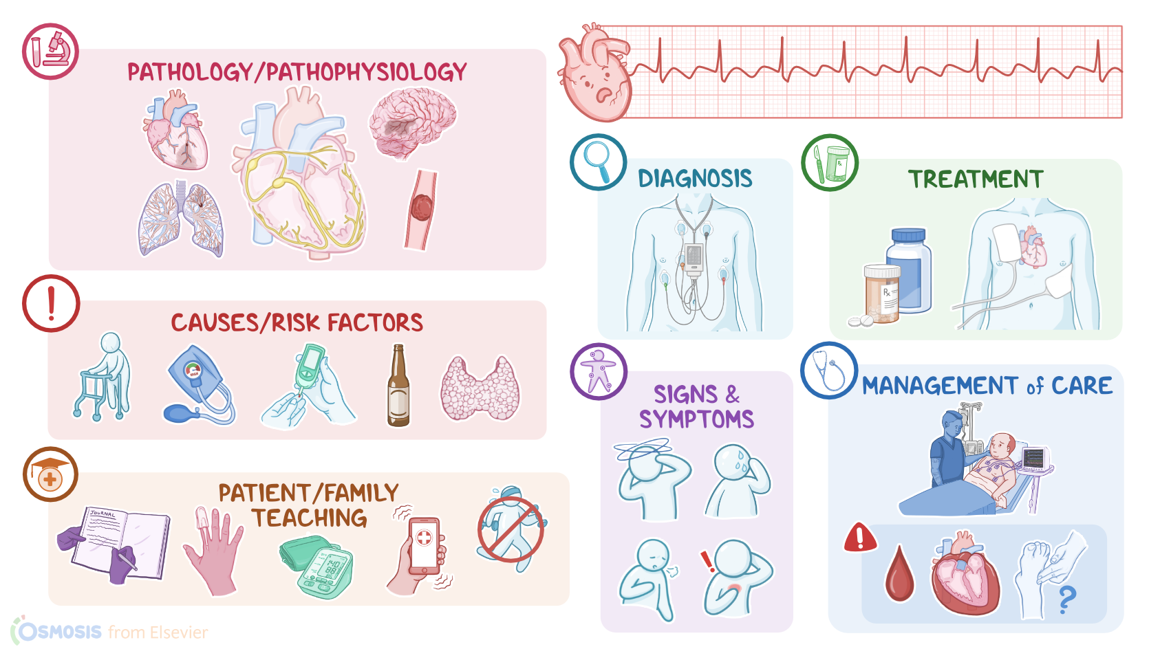
- "Critical care nursing: Diagnosis and management (10th ed.). ISBN: 978-0-443-11581-3 " Elsevier (2026)
- "Lewis’s Medical-Surgical Nursing: Assessment and Management of Clinical Problems. 12th Edition. ISBN:978-0-323-78961-5 " Elsevier
- "Heart failure and atrial flutter: a systematic review of current knowledge and practices. 8(6):4484-4496." ESC Heart Fail. (2021)
- "Stroke and thromboembolism prevention in atrial fibrillation. 106(1):10-17." Heart. (2020)
- "Prevention and Treatment of Atrial Fibrillation via Risk Factor Modification. 160:46-52." Am J Cardiol. (2021)
- "Fundamentals of nursing (10th ed.). ISBN 978-0323810340 " Elsevier (2021)
- "Saunders Comprehensive Review for the NCLEX-RN® Examination. ISBN 978-0323795302 " Saunders (2022)
- "2021 CAEP Acute Atrial Fibrillation/Flutter Best Practices Checklist. 23(5):604-610." CJEM. (2021)
No notes for this video yet
Try adding a note below